|
|
„BABEŞ-BOLYAI”
UNIVERSITY Faculty of Chemistry and
Chemical Engineering |
|


Sclerotinia sclerotiorum laccase: biochemical characterization and
applications
- PhD thesis public summary-
PhD Candidate: Augustin-Cătălin
Moţ
PhD Supervisor: Prof. Dr. Florin
Dan Irimie
|
|
„BABEŞ-BOLYAI”
UNIVERSITY Faculty of Chemistry and
Chemical Engineering |
|


Augustin-Cătălin Moţ
Sclerotinia sclerotiorum laccase: biochemical characterization and
applications
- PhD thesis -
Doctoral committee
President:
Prof. Dr. Mircea Dărăbanţu, Faculty of Chemistry and Chemical Engineering,
PhD
Supervisor: Prof. Dr. Florin Dan Irimie,
Faculty of Chemistry and Chemical Engineering,
Reviewers:
CS I Dr.
ŞTEFAN EUGEN SZEDLACSEK, Institute
of Biochemistry,
Prof. Dr.
CARMEN SOCACIU, University of
Agricultural Sciences and Veterinary Medicine,
Conf. Dr.
RADU SILAGHI-DUMITRESCU, Faculty of
Chemistry and Chemical Engineering,
|
|
|
|
|
|
|
|
|
GUVERNUL ROMÂNIEI MINISTERUL MUNCII, FAMILIEI ŞI PROTECŢIEI SOCIALE AMPOSDRU |
Fondul Social European POSDRU 2007-2013 |
Instrumente Structurale 2007-2013 |
|
OIPOSDRU |
UNIVERSITATEA BABEŞ-BOLYAI CLUJ-NAPOCA |


|
Investing in people! Ph.D. scholarship, Project co-financed by the SECTORAL OPERATIONAL PROGRAM
FOR HUMAN RESOURCES DEVELOPMENT 2007 – 2013 Priority Axis 1. "Education and training in support for
growth and development of a knowledge based society" Key area of intervention 1.5: Doctoral and post-doctoral programs in support of research. Contract nr.: POSDRU/88/1.5/S/60185 – “Innovative doctoral studies in a Knowledge Based Society”
Babeş-Bolyai University, Cluj-Napoca,
Romania |
Dedicated to my beloved wife,
Rodica and to my joyful daughter, Olga.
Table of
Contents
1.2. Overall
structure of laccases
1.21.
Architectural features of laccases
1.2.2.
C-terminus in asco-laccases
1.2.3.
Laccases with quaternary structure
1.3. Active
sites structure of laccases
1.3.1.
Type 1 copper active site
1.3.1.1.
Spectroscopic features of type 1 copper center
1.3.1.2.
Structure of type 1 copper center
1.3.1.3.
Redox potential of the type 1 copper
1.3.1.4.
Substrate binding pocket
1.4.
Catalytic mechanism of laccases
1.4.1.
Oxygen reduction to water
1.5.
Laccases purification, characterization and applications
1.5.1. Natural
sources of laccases and their physiologic roles
1.5.2.
Purification of laccases
1.5.3.
Laccases characterization
1.5.4.
Applications of laccases
1.6.
Sclerotinia sclerotiorum as laccase source candidate
1.7. Copper
complexes as models for laccase active sites
1.6.1
Model compounds for type 1 copper site
1.6.2.
Model compounds for type 2/3 copper sites
2.2.2.
Culture media and growth conditions
2.2.3.
Screening for laccase inducers
2.2.4.
Carbon and nitrogen sources
2.2.5.
Yeast extract as laccase inducer
2.2.6. Chelidonium majus extract as laccase
inducer
2.2.7.
pH and its role in laccase induction
2.2.9.
Laccase activity measurements
2.3.1.
Screening for laccase inducers
2.3.2.
Carbon and nitrogen sources as laccase regulators
2.3.3.
Yeast extracts enhance laccase production
2.3.4.
Influence of Chelidonium majus
extract upon laccase production.
2.3.5.
pH as regulator of laccase biosynthesis in Sclerotinia
sclerotiorum
Chapter 3. Isolation, purification and characterization
of S. sclerotiorum laccase
3.2.1.
Media and grow conditions
3.2.3.
Enzyme assay and protein determination
3.2.4.
Enzyme characterization
3.2.4.2.
Molecular weight determination.
3.2.6.
Mass spectrometric characterization of the laccase
3.3.1.
Culturing and laccase purification
Chapter 4. Insights into S. sclerotiorum laccase mechanisms
4.2.4.
UV-vis and fluorescence measurements
4.3.2.
Substrate-specific adduct colors
4.3.3.
ABTS binds to a Tyr residue
5.2.2.
Hemoglobin and laccase purification
5.2.3.
Enzyme kinetics measurements
5.2.4.
Pro-oxidant and antioxidant activity measurements
5.2.5.
Quercetin radical investigation by UV-vis and EPR spectroscopies
5.2.6.
Propolis extracts preparation and investigation
5.2.7.
Cyclic voltametry measurements
5.2.8.
HPLC-MS and MS investigations
5.2.9.
Folin-Ciocalteu and hemoglobin/ascorbate peroxidase activity inhibition
5.3.1.
Characterization of flavonoid substrates
5.3.2.
EPR and UV-vis detection of a species assigned as a flavonoid radical
5.3.3.
Laccase-induced prooxidant reactivity of flavonoids on hemoglobin
5.3.4.
Applications on antioxidant and pro-oxidant activities of Romanian propolis
extracts
5.3.4.1.
Antioxidant evaluation of propolis extracts
5.3.4.1.
Pro-oxidant evaluation of propolis extracts
Chapter 6. Models and theoretical approaches on laccase
active sites
6.1. Type 1
copper active site investigation
6.2. Type
2/3 copper active site investigation
List of
Figures
Figure
12. Laccase
catalytic cycle in absence (up) and presence (down) of a mediator.
Figure
40. CD spectrum of
S. sclerotiorum laccase (9.4 µM) as purified in 5 mM TAPS pH 7.8
Figure
44. ABTS-tyrosine
(black) and ABTS-laccase (grey) UV-vis spectra at pH 6.3 (25 mM MES).
Figure
62. Correlation of
the proposed QFs and the %DPPH250s for the studied propolis samples.
List of Tables
Table 1. Redox potentials of various laccases and
corresponding sequence alignments. The conserved HCH tripeptide, axial ligand
(last) are marked in bold. N.d refers to laccases whose sequence was not
determined. The pair of aminoacids marked in bold and italics is involved in
the Piontek hypothesis (see text). Unless stated (UP – uniprot database, PDB –
Protein Data Bank), the sequence codes are from GenBank. E0 is
measured vs. NHE......................... 14
Table 2. Recent cultivation conditions
and purification procedures with their yield and fold factors for several
laccase purifications from different organisms.............................................. 29
Table 3. Statistical data of Km
and kcat values for most used laccase substrates...................... 30
Table 4. Some characterization data
regarding some recently purified laccases. The references for each organism can
be found in Table 2......................................................................... 31
Table 5. Effects of various potential
laccase inducers upon the laccase activity in S. sclerotiorum................................................................................................................................... 49
Table 6. Biomass and laccase activity
variations when different carbon and nitrogen sources were used............................................................................................................................ 50
Table 7. Concentration/purification of
laccase using salt precipitation and chromatographic methods...................................................................................................................... 68
Table 8. Substrate catalytic
parameters of the purified laccase obtained by non-linear fitting model using
Origin 8............................................................................................................. 74
Table 9. Michaels-Menten parameters
for three substrates for five forms of the purified laccase obtained by
Eadie–Hofstee linearization in case of biphasic cases (Q0H2)
and non-linear fitting using GraphPad for normal curves................................................................................ 93
Table 10. Prooxidant, antioxidant,
enzymatic kinetic parameters and redox potentials of the studied compounds................................................................................................................ 108
Table 11. Floral, geographical origins
and description of the studied propolis samples......... 119
Table 12. Correlation coefficients
between several calculated parameters of the kinetic profile of DPPH bleaching
assays and some relevant IR and UV-vis absorbances......................... 121
Table 13. Distinct bands in FT-IR
spectra found in propolis extracts................................... 122
Table 14. Geographical origins and
their numbering the propolis samples taken into this section study (interaction
with hemoglobin)........................................................................... 125
Table 15. Correlation coefficients
between various antioxidant measurement methods. “DPPH” denotes the percent
decrease in DPPH absorbance in 680 seconds. GAE denotes the gallic acid
equivalents, in mg/mL. HAPX denotes the ratio between rates of ascorbate
consumption by hemoglobin and peroxide in the absence and presence of propolis,
respectively. EPR denotes the area under the EPR signal; as determined by
double integration. Details are found in Materials
and Methods............................................................................................... 130
Table 16. Several parameters regarding
binary mixture experiments................................... 133
Abbreviations
Å – ångström (1Å=10-10 m);
ABTS – a well known laccase substrate:
2,2'-azino-bis(3-ethylbenzothiazoline-6-sulphonic acid);
ANOVA – analysis of variance;
APCI – atmospheric-pressure chemical ionization;
AUC – area under the curve;
C/N – carbon to nitrogen ratio;
cAMP – cyclic adenosine monophosphate;
CAT – catalase;
CD – circular dichroism;
CER – ceruloplasmin;
CotA – the protein (with laccase activity) encoded by the gene with the same
name involved in spor coat dvelopement of Bacillus
subtilis;
CueO – copper efflux oxidase (the laccase from Escherichia coli);
CuTPP – [5, 10, 15, 20-tetrakis(N-methylpyridyl-4)porhinato]copper(II)
tetratosylate);
2,6-DMP – 2,6-dimethoxyphenol;
D4h – denotes a symmetry group;
Da – daltons;
DAD – diode array detector;
DMPO – 5,5-dimethyl-pyrroline N-oxide;
DPPH – 2,2-diphenyl-1-picrylhydrazyl;
DTT – dithyotreitol;
E○ – normal electrode potential;
EPR – electron paramagnetic resonance;
ESI – electrospray ionization;
FPLC – fast protein liquid chromatography;
(FT)IR – Fourier transformed infrared;
GAE – gallic acid equivalent;
GuHCl – guanidine hydrochloride;
HAPX – hemoglobin-ascorbate peroxidase;
Hb – hemoglobin;
HOMO – highest occupied molecular orbital;
kcat – catalytic constant (turnover number);
Km – Michaelis-Menten constant;
LAC – three domain laccase (common laccase);
LC(-MS) – liquid chromatography (-mass spectrometry);
LMCT – ligand to metal charge transfer;
LUMO – lowest unoccupied molecular orbital;
MES – 2-(N-morpholino)ethanesulfonic acid used as buffer;
MOPS – 3-(N-morpholino)propanesulfonic acid used as buffer;
NHE – normal hydrogen electrode;
nm – nanometer;
NMR – nuclear magnetic resonance;
NR – nitrite reductase;
p – the probability of obtaining a statistic test;
PAGE – polyacrylamide gel electrophoresis;
PBS – phosphate buffer saline;
PCA –Principal Component Analysis;
PDB – protein data bank;
Ph-OH – generic formula for phenolic compound;
pQF – prooxidant quercetin factor;
Q0 – dimethoxy-5-methyl-p-benzoquinone;
QF – quercetin factor;
QM/MM – quantum mechanical and molecular mechanics;
RGB – red green blue color channel;
RNS – reactive nitrogen species;
ROS – reactive oxygen species;
RP – reversed phase;
rpm – rotations per minute;
RSD – relative standard deviation;
SD – standard deviation;
SDS – sodium dodecyl sulfate;
SLAC – small laccase (one domain laccase);
SOD –superoxide dismutase;
T1Cu – type 1 copper;
T2Cu – type 2 copper;
T3Cu – type 3 copper;
TAPS – 3-[[1,3-dihydroxy-2-(hydroxymethyl)propan-2-yl]amino]propane-1-sulfonic
acid used as buffer;
TEAC – trolox equivalent antioxidant capacity;
TMB – tetramethylbenzidine;
TRIS – 2-amino-2-hydroxymethyl-propane-1,3-diol used as buffer;
U – enzymatic unit;
UV-vis – ultra violet and visible;
V – volt;
WE –working electrode;
Acknowledgements
I want to give thanks to prof. dr.
In the end I want to mention that I will remain
with wonderful memories from laboratory
6 wherever I will be in future, thus all people met there are nicely
hugged.
Aims of the thesis
The present thesis has five main objectives
which were stated in a preliminary form before the work was started and
suffered several adjustments during the work proceeded. Each of these objectives
contains several main steps which were foreseen in the research thesis project
or was established during the ongoing procedures.
- Determination of optimum conditions
considering maximum laccase activity in liquid culture of Sclerotinia
sclerotiorum
- Evaluation of
several types of liquid mediums upon laccase secretion;
- Assessing the
influence of numerous important parameters/conditions upon laccase
production by the fungus such as: pH, N and C sources, time, several
inducers;
- Evaluation of
some important physiologic relevant factors upon laccase regulation in
order to bring some insights of its physiologic role;
- Isolation, purification and
characterization of Sclerotinia sclerotiorum laccase
· Establishment of suitable protocols for laccase
isolation and purification using chromatographic and electrophoretic
facilities;
· Determination of specific activity and
biochemical properties (KM, kcat, optimum pH and
temperature, thermostability, substrate selectivity) of the purified enzyme;
· Spectral characterization of the purified
enzyme (UV-vis, CD,
- Elucidation of the mechanism of the
reactions catalyzed by the pure enzyme
· Characterization of possible reaction
intermediates and their kinetics;
· Study of enzyme – substrate interaction;
- Applications of the Sclerotinia
sclerotiorum laccase
· Establishment of protocols suitable for
evaluations of prooxidant and antioxidant activities of polyphenols and some
natural extracts;
- Theoretical and experimental studies of
model compounds for laccases
· Evaluation of reactivity towards some ligands
and laccase substrates of some copper complexes;
· Theoretical studies of laccase copper centers
concerning their reactivity and spectral behaviour.
General
Introduction
|
P |
roteins having one or more copper ions as cofactors
play very important roles in cellular metabolism of all living organisms. They
are involved in photosynthesis, oxidative phosphorylation, homeostasis of metal
ions and catabolism of many nutrients. The main reactions involving copper
proteins are electron transfer, this due to copper ability to exist in two
oxidation states Cu+ and Cu2+. Copper centers in proteins
have such a coordination sphere provided by the polypeptidic structure so that
the transition from one oxidation state to another to be thermodynamically
favorable.
The
simplest copper dependent proteins are azurins and plastocyanines, they are
usually involved in electron transfer reactions. Other more complex proteins,
with copper ions in the active sites, such as galactose oxidase, nitrite
reductase, ceruloplasmin, ascorbate reductase, bilirubin oxidase and last but
not least laccase, are involved electron transfer reactions from reduced
substrates to electron deficient molecules.
Laccase (p-diphenols: dioxigen oxidoreductase) is an
oxidoreductase (EC 1.10.3.2) with four copper ions in two active sites, which
catalyzes the oxidation of reduced substrates usually phenols or aromatic
amines, coupled with the reduction of molecular oxygen to water. Laccase is one
of the oldest enzymes ever studied, it was described for the first time by
Yoshida (1883) and categorized by Bertrand (1895) as a copper containing
oxidase. However, only in recent decades, when it was discovered that laccases
are part of the enzymatic arsenal involved in wood degradation by white rot
fungi, study of these enzymes has greatly increased. A more recent interest in
this enzyme is its involvement in the virulence of some phytopathogenic fungi,
as is the case of the present thesis.
Currently, this enzyme is the central subject of many
worldwide research groups, due to scientific curiosity and its high potential
in numerous applications in biotechnology and bioanalytical chemistry.
Summary of the content of the thesis
The
first chapter describes the most recent research on the overall structural
features of laccases as well as on the structures and properties of the active
sites, along with the currently proposed mechanisms of reaction. Laccase (p-diphenol:dioxygen oxidoreductase), one
of the oldest discovered enzymes, contains four copper ions in two active sites
and catalyzes a monoelectronic oxidation of substrates such as phenols and
their derivatives, or aromatic amines, coupled to a four-electron reduction of
dioxygen to water. The catalytic mechanism was studied for decades but is still
not completely elucidated, especially in terms of the reduction of dioxygen to
water. The key structural features of this enzyme are under research in several
groups using techniques such as X-ray diffraction, electron paramagnetic
resonance (EPR) spectroscopy, site-directed mutagenesis. The
high interest in laccases is explained by the large number of biotechnological
applications. Their distribution in nature, the physiologic role, most used
methods for purification and biochemical properties and parameters used for
their characterization are also described. Numerous applications of laccases
such as textile industry, wood processing paper
production, pharmaceutical and chemical industries and others are described.
Some biological aspects regarding Sclerotinia
sclerotiorum phytopathogenic fungus and reasons for using this organism as
laccase source are presented at the end of the chapter. In the last part of the
chapter some copper complexes used as models for laccase active sites are
discussed.
The second chapter describes the factors affecting the production of laccase from the phytopathogenic fungus Sclerotinia sclerotiorum (Lib.) de Bary. The carbon/nitrogen ratio appears to be of great importance. Rather than a simple nutrient-rich nitrogen source, yeast extract behaves as a true laccase upregulator, apparently acting via a stress pathway. Chelidonium majus extract, a known antifungal agent, acts in a similar manner. The compound(s) in the yeast extract responsible for enhancing laccase synthesis are suggested to be hydrolysable small organic molecules. Both extracts reduce biomass and sclerotia development and enhance laccase production, leading to an increase in laccase activity by one order of magnitude compared to controls. The pH of the medium, a well-known virulence regulator for this fungus, also acts as a true laccase regulator, though via a different mechanism. The effect of pH appeared to be linked to the acidification kinetics of the extracellular medium during fungal development. A number of other known laccase inducers were found to enhance laccase production at most two-fold.
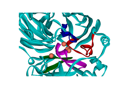
Chapter three contains information regarding the production, purification and characterization of a laccase from the phytophathogenic fungus Sclerotinia sclerotiorum. This laccase is identified by mass spectrometry with a sequence coverage of 74.9% (458/577 AA) revealing that the protein is identical or highly homologous to a predicted oxidoreductase from this species (A7EM18 in the Uniprot database); the closest homologous protein previously isolated from a fungus is the Melanocarpus albomyces, with only 35% identity. The UV-vis spectral features of this laccase classify it as a “yellow” one. The EPR spectrum nevertheless demonstrates resemblance to blue laccases – including the type 1 center not detectable in UV-vis spectra. The presence of type 3 coppers was proven by fluorescence spectrum and by 330 nm band in UV-vis. The purified laccase has an apparent molecular mass of 70 kDa and appears as a monomer. The values of KM and kcat were determined for ABTS, 2,6-dimethoxyphenol, p-phenylenediamine and guaicol and are typical of a laccase. The optimal pH value is around 4 except for ABTS, for which activity is linearly increasing with acidity. The high laccase activity in liquid culture makes Sclerotinia sclerotiorum a useful source of laccase for practical applications.
In chapter four it is provided the first evidence that the yellow laccase isolated from Sclerotinia sclerotiorum is obtained from a blue form by covalent, but nevertheless reversible modification with a polyphenolic product. Yellow laccases lack the typical blue type 1 Cu absorption band around 600 nm, but are nevertheless multicopper oxidases with laccase properties. After separating the polyphenols, a typical blue laccase is obtained. With ABTS as model substrate for this blue enzyme, a purple adduct is formed with a spectrum nearly identical to that of the 1:1 adduct of an ABTS radical and Tyr. This modification significantly increases the stability and substrate affinity of the enzyme, not by acting primarily as bound mediator, but by allosteric activation that also alters the type 1 Cu site. Thus, S. sclerotiorum yellow laccase is an intrinsically blue multi-copper oxidase that autocatalytically activates itself upon first encounter with a radical-forming aromatic substrate.
The fifth chapter contains numerous results regarding the application of the purified enzyme on antioxidant and prooxidant properties of some phenolics and propolis extracts. A transient species may be detected with UV-vis and EPR spectroscopy during turnover of a laccase with quercetin; this species is assigned as a quercetin-derived radical, based on EPR spectra as well as based on UV-vis similarities with previously reported data on a quercetyl radical obtained via a non-enzymatic route. The formation and decay of this species correlate well with the prooxidant reactivity manifested by flavonoids in the presence of laccase. An assay for the prooxidant reactivity of natural compounds is proposed based on the results reported here; this assay has the advantages of using a biologically-relevant process (hemoglobin oxidation), and of not needing added oxidizing agents such as peroxide or superoxide. Correlations, or the lack thereof, between the prooxidant parameters and the redox potentials, antioxidant capacities and lipophilicities, are analyzed. New assays for antioxidant activity of natural extracts are also described. It can be noted that the laccase employed in this study does display structural and reactivity-related similarities to a range of other proteins, which includes ceruloplasmin.
The last chapter of the thesis contains the results regarding molecular modelling of laccase active sites and the experiments describing the reactivity of some copper complexes used as models for type 2 copper sites. Laccases contain a blue mononuclear copper center known as ‘type-1’, and thought to be the primary electron acceptor from organic substrates during the catalytic cycle. A small group of laccases are also known that lack the 600 nm band and hence the blue color (“yellow laccases”). In first section it is reported the use of semiempirical (ZINDO/S-CI) calculations in order to simulate UV-vis spectral parameters for the laccase type 1 copper, attempting to assign geometrical and electronic structure elements that may control the color of this site. The ~600-nm band of the type 1 copper is confirmed to arise mainly from sulfur-to-copper charge transfer, and strong distortions allowing for its displacement by more than 200 nm and/or its dissolution are identified. In the second section some copper porphyrinates are analysed with respect to its reactivity towards some laccase substrate and some other redox active compounds. Copper porphyrinates are generally known to display a less diverse reactivity compared to their iron counterparts. It is examined a water-soluble copper porphyrinate for its ability to engage in reactions involving axial ligation to the copper or possible redox cycling. Although UV–vis spectra indicate an expected lack of reactivity, electron paramagnetic resonance spectra (EPR) reveal an unexpected wealth of changes in electronic structures at the copper, induced by potential ligands such as imidazole or nitrite, but also by seemingly unexpected candidates for ligands, such as 2,2′-azino-bis(3-ethylbenzthiazoline-6-sulphonic acid) (ABTS) and guaiacol, as well as by dithionite. An important function of many copper-containing proteins is activation of O2 and subsequent substrate oxidation. The Cu (III) oxidation state is generally considered to be less accessible because of the highly positive Cu (III)/Cu (II) redox potentials with typical amino acid ligands. In the last part it is employed density functional (DFT) calculations to explore to what extent copper (III) may be accessed in a biologically-relevant coordination environment around a mononuclear copper center, by breaking the oxygen-oxygen bond in a copper-(hydro) peroxide complex. In agreement with previous findings on copper models with related coordination patterns, the formally high-valent copper complex produced by O-O bond cleavage appears to in fact harbor both oxidizing equivalents on the ligands. The potential energy surface for such a reaction reveals that with the three-histidine binding motif at the copper, O-O bond cleavage is not impossible, but rather disfavored thermodynamically.
General
conclusions
·
Optimal
conditions under which the S. sclerotiorum
laccase can be produced were determined. The carbon and nitrogen sources and
C/N ratio appear to be of great importance for laccase production in this
fungus. Rather than a simple nutrient-rich nitrogen source, yeast extract
behaves as a true laccase inducer/upregulator, apparently acting via a stress
pathway. Chelidonium majus extract, a
known antifungal agent, acts in a similar manner. The pH of the medium, a
well-known virulence regulator for this fungus, also acts as a true laccase
regulator, though via a different mechanism. The effect of pH appears to be
linked to the acidification kinetics of the extracellular medium during fungal
development. Thus, evidence is shown that this enzyme is involved in stress
response pathways, most likely connected to virulence.
·
Sclerotinia sclerotiorum laccase has been isolated, and its catalytic
properties characterized. Notably, although this laccase can be classified as a
“yellow laccase” based on the UV-vis spectrum, the “blue” T1 center is nevertheless
observable in the EPR spectrum. The extent to which the S. sclerotiorum laccase may indeed allow definition of a new type
of laccase (neither truly “blue”, nor truly “yellow”) remains to be explored,
especially as for most yellow laccases the EPR spectra have not been reported;
should such a class be confirmed, a term such as “mixed blue-yellow” might be
appropriate.
·
Direct
evidence for an example where a blue laccase can be converted to a yellow form in vitro by covalent modification at the
T1 site, with metabolites produced by the laccase itself was provided.
Moreover, this autocatalytic modification significantly improves the structural
and catalytic properties of the enzyme. In essence, S. sclerotiorum yellow laccase is an intrinsically blue
multi-copper oxidase that has activated itself upon first encounter with a
polyphenolic substrate. A tyrosine residue was identified near the T1 site,
which may be the target of such modifications.
·
A
transient species may be detected with UV-vis and EPR spectroscopy during
turnover of a laccase with quercetin; this species is assigned as a
quercetin-derived radical, based on EPR spectra as well as based on
similarities with previously reported data. Furthermore, this species
correlates well with the prooxidant reactivity manifested by flavonoids in the
presence of laccase. An assay for prooxidant reactivity of natural compounds is
proposed based on these results, which has the advantages of using a
biologically-relevant process (hemoglobin oxidation), and of not needing added
oxidizing agents such as peroxide or superoxide. Correlations, or the lack
thereof, between the parameters obtained from this assay and redox potentials,
antioxidant capacities and lipophilicities, are discussed. It was also noted
that the laccase employed in this study does display structural and
reactivity-related similarities to a range of other proteins, which includes
the serum ceruloplasmin, and also displays reactivity similarities with
heme-containing peroxidases. In addition, a new more informative and effective
scale of antioxidant capacity is obtained by applying PCA on DPPH (2,
2-diphenyl-1-picrylhydrazyl) bleaching kinetic profiles. In order to obtain
comparable antioxidant activities, a non-dimensional parameter was generated
which is termed the quercetin factor (QF), which defines the ratio
between quercetin equivalent in mg/L of the assayed propolis sample and the
corresponding propolis concentration in mg/L. Further application of this
methodology to other botanical extracts will confirm this new method for
assessing antioxidant activity.
·
The
coordinative chemistry of copper porphyrinates may be distinctly more complex
than previously described, and that EPR but not UV-vis spectroscopy is the
method of choice for investigating this new chemistry.
·
Using
computational methods, torsion and elongation-type deformations have been
identified, which allow a “blue” tri-coordinated type 1 copper center to
apparently lose its characteristic 600-nm band responsible for its blue color both
by shifting it by more than 200 nm, and, in some cases, by decreasing the
extinction coefficients. However, DFT calculations suggest that such
distortions might also be detectable with EPR spectroscopy.
·
Unlike
in related iron or manganese complexes, high-valent states appear not to be
achievable via peroxo chemistry in copper complexes – even though O-O bond
cleavage per se appears to entail reasonably low energy barriers; this may be
interpreted to be due to a difference in redox potentials, which makes the
peroxide-derived hydroxo and oxo ligands easier to oxidize than Cu (II).
References
1)
Abdullah
J., Ahmad M., Heng L.Y., Karuppiah N., Sidek H., An optical biosensor based on immobilization of laccase and MBTH in
stacked films for the detection of catechol, Sensors 7 (2007) 2238-2250;
2)
Aboelella
N.W., Gherman B.F., Hill L.M.R., York J.T., Holm N., Young Jr. V.G., Cramer
C.J., Tolman W.B., Effects of thioether
substituents on the O2 reactivity of β-Diketiminate−Cu(I)
complexes: probing the role of the methionine ligand in copper monooxygenases,
J. Am. Chem. Soc. 128 (2006)
3445-3458;
3)
Aboelella
N.W., Kryatov S.V., Gherman B.F., Brennessel W.W., Young Jr. V.G., Sarangi R.,
Rybak-Akimova E.V., Hodgson K.O., Hedman B., Solomon E.I., Cramer C.J., Tolman
W.B., Dioxygen activation at a single
copper site: structure, bonding, and mechanism of formation of 1:1 Cu−O2
adducts, J. Am. Chem. Soc. 126 (2004)
16896-16911
4)
Adlercreutz
H., Mazur W., Phyto-oestrogens and
Western diseases, Ann. Med. 29 (1997)
95–120;
5)
Adman E.T.,
Copper protein structures, Adv.
Protein. Chem. 42 (1991) 144-197;
6)
Agrios
GN., Plant Pathology 5th ed., Elsevier, Academic Press,
7)
Akerstrom
B., Maghzal G.J., Winterbourn C.C., Kettle A.J., The lipocalin α1-microglobulin has radical scavenging activity,
J. Biol. Chem. 282 (2007)
31493–31503;
8)
Andberg
M., Hakulinen N., Auer S., Saloheimo M., Koivula A., Rouvinen J., Kruus K., Essential role of the C-terminus in
Melanocarpus albomyces laccase for enzyme production, catalytic properties and
structure, FEBS J. 276 (2009)
6285–6300;
9)
Andersen
S.O., Insect cuticular sclerotization: A
review, Insect Biochem. Mol. Biol. 40 (2010)
166-178;
10) Antonini E., Brunori M., Hemoglobin and myoglobin in their reaction with ligands: North-Holland,
11) Apak R., Guclu K., Demirata B., Ozyurek M.,
Celik S.E., Bektasoglu B., Berker K. I., Özyurt, D., Comparative evaluation of various total antioxidant capacity assays
applied to phenolic compounds with the CUPRAC assay, Molecules 12 (2007) 1496-1547;
12) Ardon O., Kerem Z., Hadar Y., Enhancement of laccase activity in liquid
cultures of the ligninolytic fungus Pleurotus ostreatus by cotton stalk extract,
J. Biotechnol. 51 (1996) 201-207;
13) Arora D.S., Rampal P., Laccase production by
some Phlebia species. J. Basic Microbiol. 42 (2002) 295-301;
14) Aruoma O.I., Assessment of potential prooxidant and antioxidant actions, JAOCS
73 (1996) 1617-1625;
15) Ashworth P., Dixon W.T., Secondary radicals in the autoxidation of hydroquinones and quinones,
J. Chem. Soc. D 9 (1972) 1130-1133;
16) Augustine A.J., Kjaergaard C., Qayyum M.,
Ziegler L., Kosman D.J., Hodgson K.O., Hedman B., Solomon E.I., Systematic perturbation of the trinuclear
copper cluster in the multicopper oxidases: the role of active site asymmetry
in itsrReduction of O2 to H2O, J. Am. Chem. Soc. 132
(2010) 6057–6067;
17)
18) Averill B., Eldredge P., General Chemistry: principles, patterns, and applications, Prentice
Hall, (2006) Chapter 23;
19) Ayse K., Beraat O., Samim S., Review of methods to determine antioxidant
capacities, Food Anal. Methods 2 (2009)
41-60;
20) Baldrian P., Fungal laccases – occurrence and properties, FEMS Microbiol. Rev.
30 (2006) 215–242;
21) Bankova V.S., Popov S.S., Marekov N.I., A study on flavonoids of propolis, J.
Nat. Prod. 46 (1983) 471-474;
22) Bankova V.S., Chemical diversity of propolis and the problem of standardization,
J. Ethnopharmacol. 100 (2005a)
114–117;
23) Bankova V.S., Recent trends and important developments in propolis research,
Evid. base Compl. Alternative Med. 2 (2005b)
29-32;
24) Banskota A.H., Tezuka Y., Adnyana I.K., Ishii
E., Midorikawa K., Matsushige K., Kadota S. Hepatoprotective
and anti Helicobacter pylori activities of constituents from Brazilian propolis,
Phytomedicine, 8 (2001) 16–23;
25) Bar-Nun N., Lev A.T., Harel E., Mayer A.M., Repression of laccase formation in Botrytis
cinerea and its possible relation to phytopathogenicity, Phytochemistry 27
(1988) 2505–2509;
26) Barreca A.M., Fabbrini M., Galli C., Gentili
P., Ljunggren S., Laccase/mediated
oxidation of a lignin model for improved delignification procedures, J.
Mol. Catal. B: Enzymatic 26 (2003)
105–10;
27) Bast A., Haenen G.R., Doelman C.J., Oxidants and antioxidants: state of the art,
Am. J. Med. 91 (1991) 2–13;
28) Bauer C.G., Kuehn A., Gajovic N., Skorobogatko
O., Holt P.J., Bruce N.C., Makower A., Lowe C.R., Scheller F.W., New enzyme sensors for morphine and codeine
based on morphine dehydrogenase and laccase, Fresenius J. Anal. Chem. 364 (1999) 179–183;
29) Bayrakçeken F., Aktas S., Toptan M., Unlugedik
A., High resolution electronic absorption
spectra of anisole and phenoxyl radical, Spectrochim. Acta 59 (2003) 135–138;
30) Beek T.A., Kuster B., Claassen F.W., Tienvieri
T., Bertaud F., Lenon G., Fungal
bio-treatment of sprucewood with Trametes versicolor for pitch control:
Influence on extractive contents, pulping process parameters, paper quality and
effluent toxicity, Biores.Technol. 98 (2007)
302–11;
31) Bento I., Martins L.O., Lopes G.G., Carrondo
M.A., Lindley P.F., Dioxygen reduction by
multi-copper oxidases; a structural perspective, Dalton. Trans. 21 (2005) 3507-3513;
32) Bent, I., Silva C.S., Chen Z., Martins L.O.,
Lindley P.F., Soares C.M., Mechanisms
underlying dioxygen reduction in laccases. Structural and modelling studies
focusing on proton transfer, BMC Struct. Biol. 10 (2010) 28-32;
33) Bertrand G., Sur la
laccase et sur le pouvoir oxydant de cette diastase, Reports of the
Paris Academy of Sciences (Paris) 120 (1895) 266–269;
34) Bernardi A.P.M., Lopez-Alarcon C., Aspee
A., Rech S., Von Poser G.L., Bride R., Lissp E. Antioxidant activity of flavonoids isolated from Hypericum ternum,
J. Chilean Chem. Soc., 52 (2007)
1326-1329;
35) Binz T., Canevascini G., Differential production of laccases in Dutch
elm disease pathogens Ophiostoma ulmi and O. novo-ulmi, Mycol. Res. 100 (1996)
1060–1064;
36) Bloom M., Van Zyl W.H., Joubert E., Botha A.,
De Villiers D., Patent number WO2006013530 (2006);
37) Boland G.J., Hall R., Index of plant hosts of Sclerotinia sclerotiorum, Canad. J. Plant
Pathol. 16 (1994) 93–108;
38) Bolton D.M., Thomma B.P.H.J., Nelson B.D., Sclerotinia sclerotiorum (Lib.) de Bary:
biology and molecular traits of a cosmopolitan pathogen, Molec. Plant
Pathol. 7 (2006) 1–16;
39) Bourbonnais R., Paice M.G., Reid I.D., Lanthier
P., Yaguchi M., Lignin oxidation by
laccase isozymes from Trametes versicolor and role of the mediator
2,2'-azinobis(3-ethylbenzthiazoline-6-sulfonate) in kraft lignin
depolymerization, App. Envir. Microbiol. 61 (1995) 1876–1880;
40) Bourbonnais R., Paice M.G., Demethylation and delignification of kraft
pulp by Trametes versicolor laccase in the presence of 2,2′-azinobis-(3-ethylbenzthiazoline-6-sulphonate)
acid, Appl. Microbiol. Biotechnol. 36 (2004)
823-827;
41) Brand-Williams W., Cuvelier M.E., Berset C., Use of a free radical method to evaluate
antioxidant activity, Lebensmittel-Wissenschaft Technol., 28 (1995) 25-30;
42) Briciu R.D., Kot-Wasik A., Namiesnik J., Sarbu
C., The lipophilicity indices of
flavonoids estimated by reversed-phase liquid chromatography using different
computation methods, J. Sep. Sci. 32(2009)
2066–2074;
43) Briciu R.D., Sarbu C., Lipophilicity of flavonoids estimated by reversed-phase high
performance thin-layer chromatography: chemically bonded plates vs. impregnated
plates with oils, animal, and human fats, Separ. Sci. Technol. 45 (2010) 1275–1285;
44) Brijwani K., Rigdon A., Vadlani P.V., Fungal laccases: production, function, and
applications in food processing, Enzyme Res. (2010), ID 149748, 10 pages;
45) Brouwers G.-J., de Vrind J.P., Corstiens P.L.,
Cornelis P., Baysse C., de Vrind-de Jong E.W., CumA, a gene encoding a multicopper oxidase, is involved in Mn2+
oxidation in Pseudomonas putida GB-1, Appl. Environ. Microbiol. 65 (1999) 1762–1768;
46) Buijs W., Comba P., Corneli D., Pritzkow H., Structural and mechanistic studies of the
copper(II)-assisted ortho-hydroxylation of benzoates by trimethylamine N-oxide,
J. Organomet. Chem. 641 (2002)
71-80;
47) Bukh C.,
48) Bulter T., Alcalde M., Sieber V., Meinhold P.,
Schlachtbauer C., Arnold F.H., Functional
expression of a fungal laccase in Saccharomyces cerevisiae by directed
evolution, Appl. Environ. Microbiol. 69 (2003) 987–995;
49) Burda S., Oleszek W., Antioxidant and antiradical activities of flavonoids, J. Agric.
Food Chem. 49 (2001) 2774–2779.
50) Burdock G.A., Review of the biological properties and toxicity of propolis, Food
Chem. Toxicol., 36 (1998) 341–363;
51) Buswell J.A., Cai Y., Chang S., Effect of nutrient nitrogen and manganese on
laccase production by L. edodes, FEMS Microbiol. Lett. 128 (1995) 81-88;
52) Calcaterra A., Galli C., Gentili P., Phenolic compounds as likely natural
mediators of laccase: A mechanistic assessment, J. Mol. Catal. B: Enzym. 51
(2008) 118–120;
53) Calle C., Schweiger A., Mitrikas G., Continuous-wave and pulse EPR study of the
copper(II) complex of N-confused tetraphenylporphyrin: direct observation of a
sigma metal-carbon bond, Inorg. Chem. 46 (2007) 1847-55;
54) Canada A.T., Giannella E., Nguyen T.D., Mason
R.P., The production of reactive oxygen
species by dietary flavonols, Free Radic. Biol. Med. 9 (1990) 441–449;
55) Cao G., Prior R.L., Anthocyanins are detected in human plasma after oral administration of
an elderberry extract, Clin. Chem. 45 (1999)
574-576;
56) Cao G., Sofic E., Prior R.L., Antioxidant and prooxidant behavior of
flavonoids: structure-activity relationships, Free Radic. Biol. Med. 22 (1997) 749–760;
57) Cessna S.G., Sears V.E., Dickman M.B., Low
P.S., Oxalic acid, a pathogenicity factor
for Sclerotinia sclerotiorum, suppresses the oxidative burst of the host plant,
The Plant Cell 2 (2000) 2191–2199;
58) Chakroun H., Mechichi T., Jesus Martinez M.,
Dhouib A., Sayadi S., Purification and
characterization of a novel laccase from the ascomycete Trichoderma atroviride:
Application on bioremediation of phenolic compounds, Process Biochem. 45 (2010) 507–513;
59) Chan T., Galati G., O'Brien P.J., Oxygen activation during peroxidase
catalysed metabolism of flavones or flavanones, Chem. Biol. Interact. 122 (1999) 15–25;
60) Chen C.C., Hwang J.K., Yang J.M., (PS)2-v2: template-based protein structure
prediction server, Bioinformatics 10 (2009)
366-379;
61) Chen Z., Durão P., Silva C.S., Pereira M.M.,
Todorovic S., Hildebrandt P., Bento I., Lindley P. F., Martins L.O., The role of Glu498 in the dioxygen
reactivity of CotA-laccase from Bacillus subtilis, Dalton. Trans. 39 (2010) 2875-2882;
62) Chernykh A., Myasoedova N., Kolomytseva M.,
Ferraroni M., Briganti F., Scozzafava A., Golovleva L., Laccase isoforms with unusual properties from the basidiomycete
Steccherinum ochraceum strain 1833, J. Appl. Microbiol. 105 (2008) 2065–2075;
63) Christenson A., Dimcheva N., Ferapontova E.E.,
Gorton L., Ruzgas T., Stoica L., Shleev S., Yaropolov A.I., Haltrich D.,
Thorneley R.N.F., Austf S.D., Direct
Electron Transfer Between Ligninolytic Redox Enzymes and Electrodes,
Electroanal. 16 (2004) 13-14;
64) Clark P.A., Solomon E.I., Magnetic circular dichroism spectroscopic definition of the
intermediate produced in the reduction of dioxygen to water by native laccase,
J. Am. Chem. Soc. 114 (1992)
1108–1110;
65) Clark K., Penner-Hahn J.E., Whittaker M.M.,
Whittaker J.W., Oxidation-state
assignments for galactose oxidase complexes from x-ray absorption spectroscopy.
Evidence for copper(II) in the active enzyme, J. Am. Chem. Soc. 112 (1990) 6433-6434;
66) Claus H., Laccases:
structure, reactions, distribution, Micron 35 (2004) 93–96;
67) Colao M.C., Garzillo A.M., Buonocore V.,
Schiesser A., Ruzzi M., Primary structure
and transcription analysis of a laccase encoding gene from the basidiomycete
Trametes trogii, Appl. Microbiol. Biotechnol. 63 (2003) 153-158;
68) Coll P.M., Fernandez-Abalos J.M., Villanueva
J.R., Santamarıa R., Perez P., Purification
and characterization of a phenoloxidase (laccase) from the lignin-degrading
Basidiomycete PM1 (CECT 2971), Appl. Environ. Microbiol. 59 (1993) 2607–2613;
69) Comba P., Knoppe S., Martin B., Rajaraman G.,
Rolli C., Shapiro B., Stork T., The
copper(II)-mediated aromatic ortho-hydroxylation: A hybrid DFT and ab initio
exploration, Chem. Eur. J. 14 (2008)
344-357;
70) Conrad L.S., Sponholz W.R., Berker O., Treatment of cork with a phenol oxidizing
enzyme, patent number US6152966 (2000);
71) Cooper C.E., Silaghi-Dumitrescu R., Rukengwa
M., Alayash A.I., Buehler P.W., Peroxidase
activity of hemoglobin towards ascorbate and urate: a synergistic protective
strategy against toxicity of Hemoglobin-Based Oxygen Carriers (HBOC),
Biochim. Biophys. Acta 1784 (2008)
1415-1420;
72) Corpet F., Multiple
sequence alignment with hierarchical clustering, Nucl. Acids Res. 16 (1988) 10881-10890;
73) Couto S.R., Toca-Herrera J.L., Laccase production at reactor scale by
filamentous fungi, Biotechnol. Adv. 25 (2007) 558–569;
74) Couto S.R., Herrera J.L.T., Industrial and biotechnological applications
of laccases: A review, Biotechnol. Adv. 24 (2006) 500–513;
75) Crestini C., Perazzini R., Saladino R., Oxidative functionalisation of lignin by
layer-by-layer immobilised laccases and laccase microcapsules, Appl. Catal.
A: General 372 (2010) 115–123;
76) Crowe J.D., Olsson S., Induction of laccase activity in Rhizoctonia solani by antagonistic
Pseudomonas fluorescens strains and a range of chemical treatments, Appl.
Envir. Microbiol. 67 (2001)
2088–2094;
77) De Souza C.G.M., Peralta R.M., Purification and characterization of the
main laccase produced by the white-rot fungus Pleurotus pulmonarius on wheat
bran solid state medium, J. Basic. Microbiol. 43 (2003) 278–286;
78) Dean J.F.D., LaFayette P.R., Rugh C., Tristram
A.H., Hoopes J.T., Eriksson K.-E.L., Merkle S.A., Laccases associated with lignifying vascular tissues. In: Lewis
N.G., Sarkanen S. (Eds.), Lignin and
Lignan Biosynthesis, ACS Symposium Series 697, American Chemical Society,
Washington, DC, (1998) p. 96;
79) Decker A., Solomon E.I., Dioxygen activation by copper, heme and non-heme iron enzymes:
comparison of electronic structures and reactivities, Curr. Opin. Chem.
Biol. 9 (2005) 152-163;
80) Ducros V., Brzozowski A.M.,
81) Dueñas M., González-Manzano S.,
González-Paramás A., Santos-Buelga C., Antioxidant
evaluation of O-methylated metabolites of catechin, epicatechin and quercetin,
J. Pharm. Biomed. Anal. 51 (2001)
443–449;
82) Dunne J., Caron A., Menu P., Alayash A.I.,
Buehler P.W., Wilson M.T., Ascorbate
removes key precursors to oxidative damage by cell-free haemoglobin in vitro
and in vivo, Biochem. J. 399 (2006)
513-24;
83) Dunne J., Svistunenko D.A., Alayash A.I.,
Wilson M.T., Cooper C.E., Reactions of
cross-linked methaemoglobins with hydrogen peroxide, Adv. Exp. Med. Biol.
471 (1999) 9-15;
84) Durao P., Chen Z., Fernandes A.T., Hildebrandt
P., Murgida D.H., Todorovic S., Pereira M.M., Melo E.P., Martins L.O., Copper incorporation into recombinant CotA
laccase from Bacillus subtilis: characterization of fully copper loaded enzymes,
J. Biol. Inorg. Chem. 13 (2008)
183–193;
85) Durman S.B., Menendez A.B., Godeas A.M., Variation in oxalic acid production and
mycelial compatibility within field populations of Sclerotinia sclerotiorum,
Soil Biol. Biochem. 37 (2005)
2180–2184;
86) Edens W.A., Goins T.Q., Dooley D., Henson J.M.,
Purification and characterization of a
secreted laccase of Gaeumannomyces graminis var. tritici., Appl. Environ.
Microbiol. 65 (1999) 3071-3074;
87) Edens W.A., Goins T.Q., Dooley D., Henson J.M.,
Purification and characterization of a
secreted laccase of Gaeumannomyces graminis var. tritici, Appl. Environ.
Microbiol. 65 (1999) 3071–3074;
88) Eggert C.,
89) Ehlinger N., Scheidt W.R., Structure and apparent reactivity of the pi-cation radical derivatives
of zinc and copper 5,10,15,20-tetra(2,6-dichlorophenyl)porphyrinate, Inorg.
Chem. 38 (1999) 1316-1321;
90) Eisenman C.H., Mues M., Weber S.E., Frases S.,
Chaskes S., Gerfen G., Casadevall A., Cryptococcus
neoformans laccase catalyses melanin synthesis from both d- and l-DOPA,
Microbiology 153 (2007) 3954–3962;
91) Elisashvili V., Kachlishvili E., Physiological regulation of laccase and
manganese peroxidase production by white-rot Basidiomycetes, J.
Biotechnol.144 (2009) 37–42;
92) Enguita F.J., Marc D.¸ Martins L.O., Grenha R.,
Henriques A.O., Lindley P.F., Carrondo M.A., Substrate and dioxygen binding to the endospore Coat laccase from
Bacillus subtilis, J. Biol. Chem. 279 (2004)
23472–23476;
93) Enguita F.J., Martins L.O., Henriques A.O.,
Carrondo M.A.,
94) Erental A., Dickman M.B., Yarden O., Sclerotial development in Sclerotinia
sclerotiorum: awakening molecular analysis of a ‘‘dormant’’ structure,
Fungal Biol. Rev. 22 (2008) 6–16;
95) Espin J.C., Soler-Rivas C., Wichers H.J., Characterization of the total free radical
scavenger capacity of vegetable oils and oil fractions using
2,2-diphenyl-1-picrylhydrazyl radical, J. Agric. Food Chem. 48 (2000) 648-656;
96) Espin J.C., Soler-Rivas C., Wichers H.J., Characterization of the total free radical
scavenger capacity of vegetables oils and oil fractions using
2,2diphenyl-1-picrylhydrazyl radical, J. Agric. Food Chem. 48 (2000) 648-656;
97) Fackler K., Kuncinger T., Ters T., Srebotnik
E., Laccase-catalyzed functionalization
with 4-hydroxy-3-methoxybenzylurea significantly improves internal bond of
particle boards, Holzforschung 62 (2008)
223-229;
98) Fakoussa R.M., Frost P.J., In vivo-decolorization of coalderived humic acids by laccase-excreting
fungus Trametes versicolor, Appl. Microbiol. Biotechnol. 52 (1999) 60–65;
99) Faraco V., Ercole C., Festa G., Giardina P.,
Piscitelli A., Sannia G., Heterologous
expression of heterodimeric laccase from Pleurotus ostreatus in Kluyveromyces
lactis, Appl. Microbiol. Biotechnol. 77 (2008) 1329–1335;
100) Fernandez-Larrea J.,
101) Ferrali M., Signorini C., Caciotti B.,
Sugherini L., Ciccoli L., Giachetti D., Comporti M., Protection against oxidative damage of erythrocyte membranes by the
flavonoid quercetin and its relation to iron chelating activity, FEBS Lett.
416 (1997) 123–129;
102) Ferraroni M.,
103) Fogliano V., Monti S.M., Musella T., Randazzo
G., Ritieni A., Formation of coloured
Maillard reaction products in a gluten-glucose model system, Food Chem. 66
(1999) 293-299;
104) Forootanfar H., Faramarzi M.A., Shahverdi A.R.,
Yazdia M.T., Purification and biochemical
characterization of extracellular laccase from the ascomycete Paraconiothyrium
variabile, Bioresour. Technol. 102 (2011)
1808-1814;
105) Fowler Z.L., Baron C.M., Panepinto J.C., Koffas
M.A.G., Melanization of flavonoids by
fungal and bacterial laccases, Yeast 28 (2011) 181-188;
106) Frei B., Higdon J.V., Antioxidant activity of tea polyphenols in vivo: evidence from animal
studies, J. Nutr. 133 (2003)
3275-3284;
107) Freire R.S., Duran N., Wang J., Kubota L.T., Laccase-based screen printed electrode for
amperometric detection of phenolic compounds, Anal. Lett. 35 (2002) 29-38;
108)
109) Fukumoto L.R., Mazza G., Assessing antioxidant and prooxidant activities of phenolic compounds,
J. Agric. Food Chem. 48 (2000)
3597-3604;
110) Galati G., Chan T., Wu B., O'Brien P.J., Glutathione-dependent generation of reactive
oxygen species by the peroxidase-catalyzed redox cycling of flavonoids,
Chem. Res. Toxicol. 12 (1999)
521–525;
111) Galati G., Moridani M.Y., Chan T.S., O’Brien
P.J., Peroxidative metabolism of apigenin
and naringenin versus luteolin and quercetin: glutathione oxidation and
conjugation, Free Radic. Biol. Med. 30 (2001) 370–382;
112) Galati G., Sabzevari O., Wilson J.X., O'Brien
P.J., Prooxidant activity and cellular effects
of the phenoxyl radicals of dietary flavonoids and other polyphenolics,
Toxicology 177 (2002) 91–104;
113) Galati G., Teng S., Moridani M.Y., Chan T.S.,
O’Brien P.J., Cancer chemoprevention and
apoptosis mechanisms induced by dietary polyphenolics, Drug Metab. Drug
Interact. 17 (2000) 311–349;
114) Galhaup C., Goller S., Peterbauer C.K., Strauss
J., Haltrich D., Characterization of the
major laccase isoenzyme from Trametes pubescens and regulation of its synthesis
by metal ions, Microbiology 148 (2002)
2159–2169;
115) Galhaup C., Haltrich D., Enhanced formation of laccase activity by the white-rot fungus Trametes
pubescens in the presence of copper, Appl. Microbiol. Biotechnol. 56 (2001) 225-232;
116) Gallaway J., Wheeldon I., Rincon R., Atanassov
P., Banta S., Barton S. C., Oxygen-reducing
enzyme cathodes produced from SLAC, a small laccase from Streptomyces
coelicolor, Biosens. Bioel. 23 (2008)
1229-1235;
117) Gamache P.H., Acworth I.N., Analysis of phytoestrogens and polyphenols
in plasma, tissue, and urine using HPLC with coulometric array detection,
Exp. Biol. Med. 217 (1998) 274-280;
118) Garavaglia S., Cambria M.T., Miglio M., Ragusa
S., Iacobazzi V., Palmieri F., D’Ambrosio C., Scaloni A., Rizzi M., The structure of Rigidoporus lignosus
laccase containing a full complement of copper ions, reveals an asymmetrical
arrangement for the T3 copper pair, J. Mol. Biol. 342 (2004) 1519–1531;
119) Gayazov R., Rodakiewicz-Nowak J., Semi-continuous production of laccase by P.
radiata in different culture media, Folia Microbiol. 41 (1996) 480-484;
120) Germann U., Muller G., Hunziker P., Lerch K., Characterization of two allelic forms of
Neurospora crassa laccase. Amino- and carboxyl-terminal processing of a
precursor, J. Biol. Chem. 263 (1988)
885–896;
121) Ghindilis A.L., Gavrilova V.P., Yaropolov A. I.,
Laccase-based biosensor for determination
of polyphenols: determination of catechols in tea, Biosens. Bioelectron.7 (1992) 127-131;
122) Giardina P., Autore F., Faraco V., Festa G.,
Palmieri G., Piscitelli A., Sannia G., Structural
characterization of heterodimeric laccases from Pleurotus ostreatus, Appl.
Microbiol. Biotechnol. 75 (2007)
1293–1300;
123) Giardina P., Faraco V., Pezzella C.,
Piscitelli A., Vanhulle S., Sannia G., Laccases:
a never-ending story, Cell. Mol. Life Sci. 67 (2010) 369–385;
124) Givaudan A., Effosse A., Faure D., Potier P.,
Bouillant M.L., Bally R., Polyphenol
oxidase in Azospirillum lipoferum isolated from rice rhizosphere: evidence for
a laccase in non-motile strains of Azospirillum lipoferum, FEMS Microbiol.
Lett. 108 (1993) 205–210;
125) Gnanamani A., Jayaprakashvel M., Arulmani M.,
Sadulla S., Effect of inducers and
culturing processes on laccase synthesis in Phanerochaete chrysosporium NCIM
1197 and the constitutive expression of laccase isozymes, Enzyme Microbiol.
Technol. 38 (2006) 1017–1021;
126) Goldman R., Claycamp R., Sweetland G.H., Sedlov
M.A., Tyurin A.V., Kisin E.R., Tyurina Y.Y., Ritov V.B., Wenger S.L., Grant
S.G., Kagan V.E., Myeloperoxidase-catalyzed
redox-cycling of phenol promotes lipid peroxidation and thiol oxidation in
HL-60 cells, Free Radic. Biol. Med. 27 (1999) 1050–1063;
127) Golz-Berner K., Walzel B., Zastrow L., Doucet
O., Cosmetic and dermatological
preparation containing copperbinding proteins for skin lightening, Patent
number WO2004017931 (2004);
128) Gomes
129) González-Forero D., Alvarez F.J., Differential postnatal maturation of GABAA,
glycine receptor, and mixed synaptic currents in renshaw cells and ventral
spinal interneurons, J. Neuroscience 25 (2005) 2010-2023;
130) Gorbacheva M., Morozova O., Shumakovich G.,
Streltsov A., Shleev S., Yaropolov A., Enzymatic
oxidation of manganese ions catalysed by laccase, Bioorg. Chem. 37 (2009) 1–5;
131) Gray H.B., Malmstrom B.G., Williams R.J., Copper coordination in blue proteins, J
Biol. Inorg. Chem. 5 (2000) 551–559;
132) Groves J.T., High-valent iron in chemical and biological oxidations, J. Inorg.
Biochem. 100 (2006) 434-47;
133) Guzy C.M., Raynor J.B., Stodulski L.P., Symons
M.C., Electron spin resonance spectra of
chromium, iron, nickel, copper and metal-free phthalocyanine reduced by sodium
in tetrahydrofuran and in hexamethylphosphoramide, J. Chem. Soc. (1969) 997-1001;
134) Haibo Z., Yinglong Z., Feng H., Peiji G.,
Jiachuan C., Purification and
characterization of a thermostable laccase with unique oxidative
characteristics from Trametes hirsuta, Biotechnol. Lett. 31 (2009) 837–843;
135) Hakulinen N., Kruus K., Koivula A., Rouvinen
J., A crystallographic and spectroscopic
study on the effect of
X-ray radiation on the crystal structure of Melanocarpus albomyces laccase, Biochem. Biophys. Res. Comm. 350 (2006) 929–934;
136) Hakulinen N., Andberg M., Kallio J., Koivula
A., Kruus K., Rouvinen J., A near atomic
resolution structure of a Melanocarpus albomyces laccase, J. Struct. Biol.
162 (2008) 29–39;
137) Halliwell B., Are polyphenols antioxidants or pro-oxidants? What do we learn from
cell culture and in vivo studies?, Arch. Biochem. Biophys. 476 (2008) 107–112;
138) Hamilton G.A., Adolf P.K., De Jersey J., DuBois
G.C., Dyrkacz G.R., Libby R.D., Trivalent
copper, superoxide, and galactose oxidase, J. Am. Chem. Soc. 100 (1978) 1899-1912;
139) Han S.K., Park H.K., A study on the preservation of meat products by natural propolis:
effect of EEP on protein change of meat products, K. J. A. Science, 37 (1995) 551–557;
140) Hanasaki Y., Ogawa S.,
141) Hao J., Song F., Huang F., Yang C., Zhang Z.,
Zheng Y., Tian, X., Production of laccase
by a newly isolated deuteromycete fungus Pestalotiopsis sp. and its
decolorization of azo dye, J. Ind. Microbiol. Biotechnol. 34 (2007) 233–240;
142) Hassett R.F., Yuan D.S., Kosman D.J., Spectral and kinetic properties of the Fet3
protein from Saccharomyces cerevisiae, a multicopper ferroxidase enzyme, J.
Biol. Chem. 273 (1998) 23274–23282;
143) Hattori M., Tsuchihara K., Noda H., Konishi H.,
Tamura Y., Shinoda T., Nakamura M., Hasegawa T., Molecular characterization and expression of laccase genes in the
salivary glands of the green rice leafhopper, Nephotettix cincticeps
(Hemiptera: Cicadellidae), Insect. Biochem. Mol. Biol. 40 (2010) 331-338;
144) Hattori M., Konishi H., Tamura Y., Konno K.,
Sogawa K., Laccase-type phenoloxidase in
salivary glands and watery saliva of the green rice leafhopper, Nephotettix
cincticeps, J. Insect Physiol. 51 (2002)1359–1365;
145) Heim K.E., Tagliaferro A.R., Bobilya D.J., Flavonoid antioxidants: chemistry,
metabolism and structure–activity relationships, J. Nutr. Biochem. 13 (2002) 572–584;
146) Heinzkill M., Bech L., Halkier T., Schneider
P., Anke T., Characterization of laccase
and peroxidase from wood-rotting fungi, Appl. Envir. Microbiol. 64 (1998) 1601-1606;
147) Hess A.V.I., Digitally enhanced thin-layer chromatography: an inexpensive, new
technique for qualitative and quantitative analysis, J. Chem. Ed. 84 (2007) 842-847;
148) Hilden K., Hakaa T.K., Lundell T., Thermotolerant and thermostable laccases,
Biotechnol. Lett. 31 (2009)
1117–1128;
149) Hirose J., Nasu M., Yokoi H., Reaction of substituted phenols with
thermostable laccase bound to Bacillus subtilis spores, Biotechnol. Lett.
25 (2003) 1609-1612;
150) Hkulinen N., Kiiskinen L.L., Kruus K., Saloheimo
M., Paananen A., Koivula A., Rouvinen J., Crystal
structure of a laccase from Melanocarpus albomyces with an intact trinuclear
copper site, Nat. Struct. Biol. 9 (2002)
601-605;
151) Hodnick W.F., Duval D.L., Pardini R.S., Inhibition of mitochondiral respiration and
cyanide-stimulated generation of reactive oxygen species by selected flavonoids,
Biochem. Pharmacol. 47 (1994)
573–580;
152)
153) Hollman P.C., van Trijp J.M., Buysman M.N., van
der Gaag M.S., Mengelers M.J., de Vries J.H., Katan M.B., Relative bioavailability of the antioxidant flavonoid quercetin from
various foods in man, FEBS Lett. 418 (1997)
152–156;
154) Huang H., Zoppellaro G., Sakurai T., A novel mixed valence form of Rhus
vernicifera laccase and its reaction with dioxygen to give a peroxide
intermediate, J. Biol. Chem. 274 (1999)
32718–32724;
155) Huang D., Ou B., Prior R.L., The chemistry behind antioxidant capacity
assays, J. Agr. Food Chem. 53 (2005)
1841-1856;
156) Huang W.T., Tai R., Hseu R.S., Huang C.T., Overexpression and characterization of a
thermostable, pH-stable and organic solvent-tolerant Ganoderma fornicatum
laccase in Pichia pastoris, Process Biochem. 46 (2011) 1469–1474;
157) Huttermann A., Mai C., Kharazipour A., Modification of lignin for the production
of new compounded materials, Appl. Microbiol. Biotechnol. 55 (2001) 387–394;
158) Hyperchem(TM) Molecular Modelling System,
Release 5.01 for Windows, Hypercube, Inc. (1998);
159) Inamo M., Kumagai H., Harada U., Itoh S.,
Iwatsuki S., Ishihara K., Takagi H.D., Electron
transfer reactions between copper(II) porphyrin complexes and various oxidizing
reagents in acetonitrile, Dalton Trans. (2004) 1703-1707;
160) Ivona J., Mirza B., Ana M., Erim B., Kajo B.,
Marica M.S., Evaluation of antioxidative
activity of Croatian propolis samples using DPPH? and ABTS2+ stable
free radical assays, Molecules 12 (2007)
1006-1021;
161) Jasprica I., Bojic M., Mornar A., Besic E.,
Bucan K., Medic-Saric M., Evaluation of
antioxidative activity of Croatian propolis samples using DPPH· and ABTS·2+
stable free radical assays, Molecules, 12 (2007) 1006-1021;
162) Jeon J.-R., Baldrian P., Murugesan K., Chang
Y.-S., Laccase-catalysed oxidations of
naturally occurring phenols: from in vivo biosynthetic pathways to green
synthetic applications, Microb. Biotechnol. 5 (2011) 318-332;
163) Johansson M., Denekamp M., Asiegbu F.O., Production and isozyme pattern of
extracellular laccase in the S and P intersterility groups of the root pathogen
Heterobasidion annosum, Mycol. Res. 103 (1999) 365–371;
164) Jones D., Ultrastructure
and composition of the cell walls of Sclerotinia sclerotiorum, Trans. Brit.
Mycol. Soc. 54 (1970) 351–360;
165) Jørgensen L.V., Madsen H.L., Thomsen M.K.,
Dragsted L.O., Skibsted L.H., Regeneration
of phenolic antioxidants from phenoxyl radicals: An ESR and electrochemical
study of antioxidant hierarchy, Free Rad. Res. 30 (1999) 207-220;
166) Josephygg P.D., Elingg T., Mason R.P., The horseradish peroxidase-catalyzed
oxidation of 3,5,3’,5’-tetramethylbenzidine, J. Biol. Chem. 257 (1982) 3669- 3675;
167) Jovanovic S.V., Steenken S., Hara Y., Simic
M.G., Reduction potentials of flavonoid
and model phenoxyl radicals. Which ring in flavonoids is responsible for
antioxidant activity?, J. Chem. Soc. Perkin Trans. 2 (1996) 2497-2504;
168) Jung H., Xu F., Li K., Purification and characterization of laccase from wood-degrading fungus
Trichophyton rubrum. LKY-7, Enzyme Microb. Technol. 30 (2002) 161-168;
169) Junghanns C., Pecyna M.J., Bohm D., Jehmlich
N., Martin C., von Bergen M., Schauer F., Hofrichter M., Schlosser D., Biochemical and molecular genetic
characterisation of a novel laccase produced by the aquatic ascomycete Phoma
sp. UHH 5-1-03, Appl. Microbiol. Biotechnol. 84 (2009) 1095-105;
170) Justino G.C., Santos M.R., Canário S., Borges
C., Florêncio M.H., Mira L., Plasma
quercetin metabolites: structure−antioxidant activity relationships,
Arch. Biochem. Biophys. 432 (2004)
109–121;
171) Kallio J.P., Gasparetti C., Andberg M., Boer
H., Koivula A., Kruus K., Rouvinen J.,
172) Kamachi T., Kihara N., Shiota Y., Yoshizawa K.,
Computational exploration of the
catalytic mechanism of dopamine beta-monooxygenase: modeling of its mononuclear
copper active sites, Inorg. Chem. 44 (2005)
4226-4236;
173) Kaneko S., Cheng M., Murai H., Takenaka S.,
Murakami S., Aoki K., Purification and
characterization of an extracellular laccase from Phlebia radiata strain
BP-11-2 that decolorizes fungal melanin, Biosci. Biotechnol. Biochem. 73 (2009) 939–942;
174) Kartal M., Yıldız S., Kaya S., Kurucu
S., Topçu G., Antimicrobial activity of
propolis samples from two different regions of Anatolia, J. Ethnopharmacol.
86 (2003) 69-73;
175) Kataoka K., Sugiyama R., Hirota S., Inoue M.,
Urata K., Minagawa Y., Seo D., Sakurai T., Four-electron
reduction of dioxygen by a multicopper oxidase, CueO, and roles of Asp112 and
Glu506 located adjacent to the trinuclear copper center, J. Biol. Chem.,
284 (2009) 14405–14413;
176) Katircioglu H., Mercan N., Antimicrobial activity and chemical compositions of Turkish propolis
from different region, Afric. J. Biotechnol. 5 (2006) 1151-1153;
177) Kiiskinen L.-L., Saloheimo M., Molecular cloning and expression in
Saccharomyces cerevisiae of a laccase gene from the ascomycete Melanocarpus
albomyces, Appl. Environ. Microbiol. 70 (2004) 137–144;
178) Kim H., Chen C., Kabbage M., Dickman M.B., Identification and characterization of
Sclerotinia sclerotiorum NADPH oxidases, Appl. Environ. Microbiol. 77 (2011) 7721-7729;
179) Kitajima N., Moro-oka Y., Copper-Dioxygen Complexes. Inorganic and Bioinorganic Perspectives,
Chem. Rev. 94 (1994) 737-757.
180) Klinman J.P., Mechanisms whereby mononuclear copper proteins functionalize organic
substrates, Chem. Rev. 96 (1996)
2541–2562;
181) Klonowska A., Gaudin C., Fournel A., Asso M.,
Le Petit J., Giorgi M., Characterization
of a low redox potential laccase from the basidiomycete C30, Eur. J.
Biochem. 269 (2002) 6119–6125;
182) Koren E., Kohen R., Ginsburg I., Polyphenols enhance total oxidant-scavenging
capacities of human blood by binding to red blood cells, Exp. Biol. Med.
(Maywood) 235 (2010) 689-699.
183) Kruus K., Kiiskinen L.-L., Saloheimo M.,
Hakulinen N., Rouvinen J., Paananen A., Linder M., Viikari L., Purification and characterisation of a novel
laccase from the ascomycete Melanocarpus albomyces, Appl. Microbiol.
Biotechnol., 59 (2002) 198-204;
184) Kumar S.V.S., Phale P.S., Durani S., Wangikar
P.P., Combined sequence and structure
analysis of the fungal laccase family, Biotechnol. Bioeng. 83 (2003) 386–394;
185) Kunamneni A., Camarero S., García-Burgos C.,
Plou F.J., Ballesteros A., Alcalde M., Engineering
and Applications of fungal laccases for organic synthesis, Microb. Cell.
Fact. 7 (2008a) 1-17;
186) Kunamneni A., Plou F.J., Ballesteros A.,
Alcalde M., Laccases and their
applications: a Patent review, Recent. Pat. Biotechnol. 2 (2008b) 10-24;
187) Kyritsis P., Messerchmidt A., Huber R., Salmon
G.A., Sykes A.G., Pulse-radiolysis
studies on the oxidised form of the multicopper enzyme ascorbate oxidase:
evidence for two intramolecular electron transfer steps, J. Chem. Soc.
Dalton Trans. (1993) 731–735;
188) Lad L., Mewies M., Basran J., Scrutton N.S.,
Raven E.L., Role of histidine 42 in
ascorbate peroxidase. Kinetic analysis of the H42A and H42E variants, Eur.
J. Biochem. 269 (2002) 3182-3192;
189) Lam H.S., Proctor A., Howard L., Cho M.J., Rapid fruit extracts antioxidant capacity
determination by fourier transform infrared spectroscopy, J. Food Science
70 (2005) 545-549;
190) Lang G., Cotteret J., Hair dye composition containing a laccase, (L'Oreal, Fr.). Int.
Pat. Appl. Patent number WO9936036, (1999);
191) Larsson S., Cassland P., Jonsson L.J., Development of a Saccharomyces cerevisiae
strain with enhanced resistance to phenolic fermentation inhibitors in
lignocellulose hydrolysates by heterologous expression of laccase, Appl.
Envir. Microbiol. 67 (2001)
1163–1170;
192) Leatham G.F., Stahmann M.A., Studies on the laccase of Lentinus edodes:
specificity, localization and association with the development of fruiting
bodies, J. Gen. Microbiol. 125 (1981)
147-157;
193) Lee S.K., George S.D., Antholine W.E., Hedman
B., Hodgson K.O., Solomon E.I., Nature of
the intermediate formed in the reduction of O2 to H2O at the trinuclear copper
cluster active site in native laccase, J. Am. Chem. Soc. 124 (2002) 6180–6193;
194) Lee D.Y., Chang G.D., Electrolytic reduction: modification of proteins occurring in
isoelectric focusing electrophoresis and in electrolytic reactions in the
presence of high salts, Anal. Chem. 81 (2009) 3957–3964;
195) Lee E.R., Kang G.H., Cho S.G., Effect of flavonoids on human health: old
subjects but new challenges, Recent Pat. Biotechnol. 1 (2007) 139-150;
196) Lee M.J., Wang Z.Y., Li H., Chen L., Sun Y.,
Gobbo S., Balentine D.A., Yang C.S., Analysis
of plasma and urinary tea polyphenols in human subjects, Cancer Epidem.
Biomar. Prev. 4 (1995) 393-399;
197) Lee-Hilz Y.Y., Boerboom A.M., Westphal A.H.,
Berkel W.J., Aarts J.M., Rietjens I.M., Pro-oxidant
activity of flavonoids induces EpRE-mediated gene expression, Chem. Res.
Toxicol. 19 (2006) 1499-1505;
198) Leontievsky A., Myasoedova N., Pozdnyakova N.,
Golovleva L., `Yellow' laccase of Panus
tigrinus oxidizes non-phenolic substrates without electron-transfer mediators,
FEBS Lett. 413 (2006) 446-448;
199) Leontievsky A.A., Myasoedova N.M., Baskunov
B.P., Pozdnyakova N.N., Vares T., Kalkkinen N., Hatakka A.I., Golovleva L.A., Reactions of blue and yellow fungal
laccases with lignin model compounds, Biochemistry (Mosc.) 64 (1999) 1150–1156;
200) Leontievsky A.A., Vares T., Lankinen P.,
Shergill J.K., Pozdnyakova N.M., Myasoedova, Kalkkinen N., Golovleva L.A.,
Cammack R., Thurston C.F., Hatakka A., Blue
and yellow laccases of ligninolytic fungi, FEMS Microbiol. Lett. 156 (1997) 9–14.
201) Levasseur A., Saloheimo M., Navarro D., Andberg
M., Pontarotti P., Kruus K., Record E., Exploring
laccase-like multicopper oxidase genes from the ascomycete Trichoderma reesei:
a functional, phylogenetic and evolutionary study, BMC Biochem. 11 (2010) 32;
202) Lindley P.F., Handbook of Metalloproteins, Marcel Dekker, Inc.,
203) Liptak M.D., Fleischhacker A.S., Matthews R.G.,
Telser J., Brunold T.C., Spectroscopic and
computational characterization of the base-off forms of cob(II)alamin, J.
Phys. Chem. B 113 (2009) 5245-5254;
204) Liptak M.D., Brunold T.C., Spectroscopic and computational studies of Co1+cobalamin: spectral and
electronic properties of the "superreduced" B12 cofactor, J. Am.
Chem. Soc. 128 (2006) 9144-9156;
205) Lisov A.V., Zavarzina A.G., Zavarzin A.A.,
Leontievsky A.A., Laccases produced by
lichens of the order Peltigerales, FEMS Microbiol. Lett. 275 (2007) 46–52;
206) Litthauer D., Jansen van Vuuren M., van Tonder
A., Wolfaardt F.W., Purification and
kinetics of a thermostable laccase from Pycnoporus sanguineus (SCC 108),
Enzyme Microb. Technol. 40 (2007)
563–568;
207) Liu L., Tewari R.P., Williamson P.R., Laccase protects Cryptococcus neoformans
from antifungal activity of alveolar macrophages, Infect. Immun. 67 (1996) 6034–6039;
208) Liu Y.T., Lu B.N., Xu L.N., Yin L.H., Wang
X.N., Peng J.Y., Liu K.X., The
antioxidant activity and hypolipidemic activity of the total flavonoids from
the fruit of Rosa laevigata, Michx. Nat. Sci. 3 (2010) 175–783;
209) Lo S.C., Ho Y.S., Buswell J.A., Effect of phenolic monomers on the
production of laccases by the edible mushroom Pleurotus sajor-caju, and partial
characterization of a major laccase component, Mycologia 93 (2001) 413-421;
210) Lomascolo A., Record E, Herpoël-Gimbert I.,
Delattre M., Robert J.L., Georis J., Dauvrin T., Sigoillot J.C., Asther M., Overproduction of laccase by a monokaryotic
strain of Pycnoporus cinnabarinus using ethanol as inducer, J. Appl.
Microbiol. 94 (2003) 618–624;
211) Lotito S.B, Frei B., Consumption of flavonoid-rich foods and increased plasma antioxidant
capacity in humans: cause, consequence, or epiphenomenon?, Free Radic.
Biol. Med. 41 (2006) 1727–1746;
212) Luna-Acosta A., Rosenfeld E., Amari M.,
Fruitier-Arnaudin I., Bustamante P., Thomas-Guyon H., First evidence of laccase activity in the Pacific oyster Crassostrea
gigas, Fish Shellfish Immunol. 28 (2010)
719-726;
213) MacDonald-Wicks L.K., Wood L.G., Garg M.L., Methodology for the determination of
biological antioxidant capacity in vitro: a review, J. Sci. Food Agr. 86 (2006) 2046-2056;
214) Maceiras R., Rodriguez C.S., Sanroman A., Influence of several activators on the
extracellular laccase activity and in vivo decolourization of poly R-478 by
semi-solid-state cultures of Trametes versicolor, Acta Biotechnol. 21 (2001) 255–264;
215) Machczynski M.C., Vijgenboom E., Samyn B.,
Canters G.W., Characterization of SLAC: A
small laccase from Streptomyces coelicolor with unprecedented activity,
Prot. Sci. 13 (2004) 2388–2397;
216) Machonkin T.E., Quintanar L., Palmer A.E.,
Hassett R., Severance S., Kosman D.J., Solomon E.I., Spectroscopy and reactivity of the type 1 copper site in Fet3p from
Saccharomyces cerevisiae: Correlation of structure with reactivity in the
multicopper oxidases, J. Am. Chem. Soc. 123 (2001) 5507–5517;
217) Mackiewicz A., Ratajczak W., Principal Component Analysis (PCA),
Comp. Geosci., 19 (1993) 303-342;
218) Maeta K., Nomur W., Takashume Y., Izawa S.,
Inove Y., Green tea polyphenols fuction
as prooxidants to active oxidative-stress-responsive transcription factors in
yeasts, Appl. Environ. Microbiol. 73 (2007)
572–580;
219) Majeau J.A., Brar S.K., Tyagi R.D., Laccases for removal of recalcitrant and
emerging pollutants, Biores. Technol. 101 (2010) 2331–2350;
220) Malkin R., Malmstrom B.G., Vanngard T., Spectroscopic differentiation of the
electron-accepting sites in fungal laccase, Eur. J. Biochem. 10 (1969) 324–329;
221) Malmstrom B.G., Rack-induced bonding in blue-copper proteins, Eur. J. Biochem. 223
(1994) 711-718;
222) Manach C., Williamson G., Morand C., Scalbert
A., Remesy C., Bioavailability and
bioefficacy of polyphenols in humans. I. Review of 97 bioavailability studies,
Am. J. Clin. Nutr. 81 (2005)
230–242;
223) Mansur M., Suárez T., González A.E., Differential gene expression in the laccase
gene family from basidiomycete I-62 (CECT 20197), App. Envir. Microbiol. 64
(1998) 771–774;
224) Manthey J.A., Fourier transform infrared spectroscopic analysis of the
polymethoxylated flavone content of orange oil residues, J. Agr. Food Chem.
54 (2006) 3215-3218;
225) Mantovani T.R.D., Linde G.A., Colauto N.B., Effect of the addition of nitrogen sources
to cassava fiber and carbon-to-nitrogen ratios on Agaricus brasiliensis growth,
Canad. J. Microbiol. 53 (2007)
139-143;
226) Marcucci M.C., Propolis: chemical composition, biological properties and therapeutic
activity, Apidology 26 (1995)
83-99;
227) Markham K.R., Mitchell K.A., Wilkins A.L.,
Daldy J.A., Lu Y., HPLC and GC-MS
identification of the major organic constituents in New Zealand propolis,
Phytochemistry 42 (1996) 205-211;
228) Martins L.O., Soares C.M., Pereira M.M.,
Teixeira M., Costa T., Jones G.H., Henriques A.O., Molecular and biochemical characterization of a highly stable bacterial
laccase that occurs as a structural component of the Bacillus subtilis
endospore coat, J. Biol. Chem. 277 (2002)
18849–18859;
229) Marukawa S., Funakawa S., Satomura Y., Role of sclerin on morphogenesis in
Sclerotinia sclerotiorum de Bary (including S. libertiana Fuckel), Agr.
Biol. Chem. 39 (1975) 645-650;
230) Matera I., Gullotto A., Tilli S., Ferraroni M.,
Scozzafava A., Briganti F., Crystal structure of the blue multicopper oxidase
from the white-rot fungus Trametes trogii complexed with ptoluate, Inorgan.
Chim. Acta 361 (2008) 4129–4137.
231) Mayera A.M., Staples R.C., Laccase: new functions for an old enzyme, Phytochemistry 60 (2002) 551–565;
232) Melníka M., Kabešová M., Cu(III) coordination compounds: classification and analysis of
crystallographic and structural data, J. Coord. Chem. 50 (2000) 323- 328
233) Melo E.P., Fernandes A.T., Durao P., Martins
L.O., Insight into stability of CotA
laccase from the spore coat of Bacillus subtilis, Biochem. Soc. Trans. 35 (2007) 1579-82;
234) Mendonc R., Ferraz A., Kordsachia O., Koch G., Cellular UV microspectrophotometric
investigations on pine wood (Pinus taeda and Pinus elliottii) delignification
during biopulping with Ceriporiopsis subvermispora (Pilat) Gilbn. & Ryv.
and alkaline sulfite/anthraquinone treatment, Wood Sci. Technol. 38 (2004) 567–75;
235) Menga F., Zuoa G., Haob X., Wangb G., Xiaob H.,
Zhangb J., Xua G., Antifungal activity of
the benzo[c]phenanthridine alkaloids from Chelidonium majus Linn against
resistant clinical yeast isolates, J. Ethnopharmacol. 125 (2009) 494–496;
236) Messerschmidt A., Multi-copper Oxidases, World Scientific,
237) Mimmi M.C., Gullotti M., Santagostini L.,
Battaini G., Monzani E., Pagliarin R., Zoppellaro G., Casella L., Models for biological trinuclear copper
clusters. Characterization and enantioselective catalytic
oxidation of catechols by the copper(II) complexes of a chiral ligand derived
from (S)-(−)-1,1′-binaphthyl-2,2′-diamine, Dalton Trans. (2004) 2192-2201;
238) Min K.L., Kim Y.H., Kim Y.W., Jung H.S., Hah
Y.C., Characterization of a novel laccase
produced by the wood-rotting fungus Phellinus ribis, Arch. Biochem.
Biophys. 392 (2001) 279–286;
239) Min K., Kim Y.H., Kim Y.W., Jung H.S., Hah
Y.C., Characterization of a novel laccase
produced by the wood-rotting fungus Phellinus ribis, Arch. Biochem.
Biophys., 392 (2001) 279-286;
240) Minussi R.C., Pastore G.M., Duran N., Laccase induction in fungi and laccase/N-OH
mediator systems applied in paper mill effluent, Bioresour. Technol. 98 (2007) 158–164;
241) Mirica L.M., Ottenwaelder X., Stack T.D., Structure and spectroscopy of
copper-dioxygen complexes, Chem. Rev. 104 (2004) 1013-1045;
242) Miyataka H., Nishiki M., Matsumoto H., Fujimoto
T., Matsuka M., Satoh T., Evaluation of
Brazilian and Chinese propolis by enzymatic and physicochemical methods,
Biol. Pharm. Bulletin, 20 (1997)
496–501;
243) Mizuno M., Food
packaging materials containing propolis as a preservative, Japanese Patent
No. JP Ol 243 974 [89 243 974] (1989);
244) Mladěnka P., Zatloukalová L., Filipský T.,
Hrdina R., Cardiovascular effects of
flavonoids are not caused only by direct antioxidant activity, Free Radic.
Biol. Med. 15 (2010) 963–75.
245) Morozova O.V., Shumakovich G.P., Gorbacheva
M.A., Shleev S.V., Yaropolov A.I., "Blue"
laccases, Biochemistry (Mosc.) 72 (2007)
1136–1150;
246) Morozova O.V., Shumakovich G.P., Shleev S.V.,
Yaropolov Y.I., Laccase-mediator systems and
their applications: A review, App. Biochem. Microb. 43 (2007) 523-535;
247) Moţ A.C., Damian G., Sarbu C.,
Silaghi-Dumitrescu R., Redox reactivity
in propolis: direct detection of free radicals in basic medium and interaction
with hemoglobin, Redox Rep. 14 (2009)
267-274;
248) Moţ A.C., Soponar F., Sârbu C., Multivariate analysis of reflectance spectra
from propolis: geographical variation in Romanian samples, Talanta 81 (2010a) 1010-1015;
249) Moţ A.C., Kis Z., Svistunenko D.A, Damian
G., Silaghi-Dumitrescu R., Makarov S.V., 'Super-reduced'
iron under physiologically-relevant conditions, Dalton Trans. 39 (2010b) 1464-1466;
250) Moţ A.C., Silaghi-Dumitrescu R., Sarbu C.,
Rapid and effective evaluation of the
antioxidant capacity of propolis extracts using DPPH bleaching kinetic
profiles, FT-IR and UV–vis spectroscopic data, J. Food Comp. Anal. 24 (2011) 516–522;
251) Moţ A.C., Pârvu M., Damian G., Irimie
F.D., Darula Z., Medzihradszky K.F., Brem B., Silaghi-Dumitrescu R., A “yellow” laccase with “blue” spectroscopic
features, from Sclerotinia sclerotiorum, Process Biochem.47 (2012) 968–975;
252) Mougin C., Boyer F.D., Caminade E., Rama R., Cleavage of the diketonitrile derivative of
the herbicide isoxaflutole by extracellular fungal oxidases, J. Agr. Food
Chem. 48 (2000) 4529–4534;
253) Murugesan K., Chang Y.Y., Kim Y.M., Jeon J.R.,
Kim E.J., Chang Y.S., Enhanced
transformation of triclosan by laccase in the presence of redox mediators, Water
Res. 44 (2010) 298-308;
254) Nakagawa H.K., Hasumi J.T., Nagai K., Wachi M.,
Generation of hydroxide peroxide
primarily contributes to the induction of Fe(II)-dependent apoptosis in Jurkat
cells by (−)-epigallocatechin-3-gallate, Carcinogenesis 25 (2004) 1567–1574;
255) Nakamura K., Go N., Function and molecular evolution of multicopper blue proteins,
Cell. Mol. Life Sci. 62 (2005)
2050–2066;
256) Neifar M., Jaouani A., Ellouze-Ghorbel R.,
Ellouze-Chaabouni S., Purification,
characterization and decolourization ability of Fomes fomentarius laccase
produced in solid medium, J. Mol. Cat. B: Enz. 64 (2010) 68–74;
257) Ness A.R., Powles J.W., Fruit and vegetables and cardiovascular disease: a review, Int. J.
Epidemiol. 26 (1997) 1–13;
258) Ng T.B., Wang H.X, A homodimeric laccase with unique characteristics from the yellow
mushroom Cantharellus cibarius, Biochem. Biophys. Res. Commun. 313 (2004) 37–41;
259) Niebisch C.H., Malinowski A.K., Schadeck R.,
Mitchell D.A., Kava-Cordeiro V., Paba J., Decolorization
and biodegradation of reactive blue 220 textile dye by Lentinus crinitus
extracellular extract, J. Hazard. Mater. 180 (2010) 316–322;
260) Nijveldt R.J., van Nood E., van Hoorn D.E.C.,
Boelens P.G., van Norren K., van Leeuwen P.A.M., Flavonoids: a review of probable mechanisms of action and potential
applications, Am. J. Clin. Nutr. 74 (2001)
418–425;
261) Nun N.B., Lev A.T., Harel E., Mayer A.M., Repression of laccase formation in Botrytis
cinerea and its possible relation to phytopathogenicity, Phytochemistry 27
(1988) 2505-2509;
262) Ohshima H., Yoshie Y., Auriol S.,
263) O'Malley D.M., Whetten R., Bao W., Chen C.L.,
Sederoff R.R., The role of of laccase in
lignification, Plant J. 4 (1993)
751–757;
264) Osawa T., Shibamoto T., Analysis of free malonaldehyde formed in lipid peroxidation systems via
a pyrimidine derivative, J. Am. Oil Chem. Soc. 69 (1992) 466-468;
265) Osiadacz J., Adhami A.J.H., Bjraszewska D.,
Fischer P., Peczyniska C.W., On the use of
Trametes versicolor laccase for the conversion of 4-methyl-3-hydroxyanthranilic
acid to actinocin chromophore, J. Biotechnol. 72 (1999) 141–149;
266) Osma J.F., Saravia V., Herrera J.L.T., Couto S.
R., Mandarin peelings: The best carbon
source to produce laccase by static cultures of Trametes pubescens,
Chemosphere 67 (2007) 1677–1680;
267) Ozcelik B., Lee J.H., Min D.B. Effects of light, oxygen, and pH on the
absorbance of 2,2-diphenyl-1-picrylhydrazyl, J. Food Sci. 68 (2003) 487-490;
268) Paganga G.,
269) Palmer A.E., Szilagyi R.K., Cherry J.R., Jones
A., Xu F., Solomon E.I., Spectroscopic
characterization of the Leu513His variant of fungal laccase: effect of increased
axial ligand interaction on the geometric and electronic structure of the type
I copper site, Inorg. Chem. 42 (2003)
4006-4017;
270) Palmer A.E., Randall D.W., Xu F., Solomon E.I.,
Spectroscopic studies and electronic
structure description of the high potential type 1 copper site in fungal
laccase: Insight into the effect of the axial ligand, J. Am. Chem. Soc. 121
(1999) 7138–7149;
271) Palmieri G., Cennamo G., Faraco V., Amoresano
A., Sannia G., Giardina P., Atypical laccase
isoenzymes from copper supplemented Pleurotus ostreatus cultures, Enzyme.
Microb. Technol. 33 (2003) 220–230;
272) Palmieri G., Giardina P., Bianco C.,
Scaloni A., Capasso A., Sannia G., A
novel white laccase from Pleurotus ostreatus, J. Biol. Chem. 272 (1997)
31301–31307;
273) Palonen H., Saloheimo M., Viikari L., Kruus K.,
Purification, characterization and
sequence analysis of a laccase from the ascomycete Mauginiella sp., Enzyme
Microb. Technol. 33 (2003) 854–862;
274) Pârvu M., Pârvu A.E., Antifungal plant extracts. In: Science
against microbial pathogens: communicating current research and technological
advances, A Méndez-Vilas (Ed.) (2011)
p. 1055-1062.
275) Pârvu M., Pârvu A.E., Crăciun C.,
Barbu-Tudoran L., Tămaş M., Antifungal
activities of Chelidonium majus extract on Botrytis cinerea in vitro and
ultrastructural changes in its conidia, J. Phytopathol. 156 (2008) 550–552;
276) Pena R.C., Propolis
standardization: a chemical and biological review, Cien. Invest. Agr., 35 (2008) 11-20;
277) Pereira L., Coelhoa A.V., Viegas C.A., Santos
M.M.C., Robalo M.P., Martins L.O., Enzymatic
biotransformation of the azo dye Sudan Orange G with bacterial CotA-laccase,
J. Biotechnol. 139 (2009) 68–77;
278) Pereira P., Bastos C., Tzanov T., Cavaco-Paulo
A., Guebitz G.M., Environmentally friendly
bleaching of cotton using laccases, Environ. Chem. Lett. 3 (2005) 66–69;
279) Periasamy R., Palvannan T., Optimization of laccase production by
Pleurotus ostreatus IMI 395545 using the Taguchi DOE methodology, J. Basic
Microbiol. 50 (2010) 548-556;
280) Pezet R., Pont V., Hoang-Van K., Evidence for oxidative detoxication of
pterostilbene and resveratrol by a laccase-like stilbene oxidase produced by
Botrytis cinerea, Physiol. Mol. Plant Pathol. 39 (1991) 441–450;
281) Pezet R., Pont V., Hoang-Van K., Enzymatic detoxication of stilbenes by
Botrytis cinerea and inhibition by grape berries proanthrocyanidins. In:
Verhoeff K., Malathrakis N.E., Williamson B., (eds), Recent Advances in Botrytis Research, Pudoc Scientific, Wageningen,
(1992) p. 87–92;
282) Pezet R., Purification
and characterization of a 32-kDa laccase-like stilbene oxidase produced by
Botrytis cinerea, FEMS Microbiol. Lett. 167 (1998) 203–208;
283) Piontek K., Antorini M., Choinowski T., Crystal structure of a laccase from the
fungus Trametes versicolor at 1.90-Å resolution containing a full complement of
coppers, J. Biol. Chem. 277 (2002)
37663–37669;
284) Piscitelli A., Giardina P., Lettera V.,
Pezzella C., Sannia G., Faraco V., Induction
and transcriptional regulation of laccases in fungi, Curr. Genet. 12 (2011) 104-112;
285) Popova T.V., Aksenova N.V., Complexes of copper in unstable oxidation
states, Russ. J. Coord. Chem. 29 (2003)
743-745;
286) Popp R., Schimmer O., Induction of sister-chromatid exchanges (SCE), polyploidy, and
micronuclei by plant flavonoids in human lymphocyte cultures. A comparative
study of 19 flavonoids, Mutat. Res. 246 (1991) 205–213;
287) Pozdnyakova N., Rodakiewicz-Nowak J.,
Turkovskaya O.V., Haber J., Oxidative
degradation of polyaromatic hydrocarbons and their derivatives catalyzed
directly by the yellow laccase from Pleurotus ostreatus D1, J. Mol. Catal.
B: Enzym. 41 (2006) 8-15;
288) Pozdnyakova N.N., Rodakiewicz-Nowak J.,
Turkovskaya O.V., Catalytic properties of
yellow laccase from Pleurotus ostreatus D1, J. Mol. Catal. B: Enzym. 30 (2004) 19–24;
289) Pozdnyakova N.N., Turkovskaya O.V., Yudina
E.N., Rodakiewicz-Nowak J., Yellow
laccase from the fungus Pleurotus ostreatus D1: purification and
characterization, Appl. Biochem. Microbiol. 42 (2006) 56–61;
290) Prior R.L., Wu X., Schaich K., Standardized methods for the determination
of antioxidant capacity and phenolics in foods and dietary supplements, J.
Agric. Food Chem. 53 (2005)
4290–4302;
291) Procházková D.,
292) Prytzyk E., Dantas A.P., Salomao K., Pereira
A.S., Bankova V.S., De Castro S.L., Aquino Neto F.R., Flavonoids and trypanocidal activity of Bulgarian propolis, J.
Ethnopharmacol., 88 (2003) 189-193;
293) Purdy L.H., Sclerotinia
sclerotiorum: history, diseases and symptomatology, host range, geographic
distribution, and impact, Phytopathology 69 (1979) 875-880;
294) Quintanar L., Stoj C., Taylor A.B., Hart P.J.,
Kosman D.J., Solomon E.I., Shall we
dance? How a multicopper oxidase chooses its electron transfer partner, Acc.
Chem. Res. 40 (2007) 445-452;
295) Quintanar L., Stoj C., Wang T.P., Kosman D.J.,
Solomon E.I., Role of aspartate 94 in the
decay of the peroxide intermediate in the multicopper oxidase Fet3p,
Biochemistry 44 (2005) 6081–6091;
296) Raaman N, Phytochemical
Techniques, New India Publishing Agency, New Delhi, India (2006);
297) Randall D.W., DeBeer George S. Hedman B.,
Hodgson K.O., Fujisawa K., Solomon E.I., Spectroscopic
and rlectronic structural studies of blue copper model complexes. 1.
perturbation of the thiolate−Cu bond, J. Am. Chem. Soc. 122 (2000) 11620-11631
298) Randall D.W., DeBeer George S., Holland P.L.,
Hedman B., Hodgson K.O., Tolman W.B., Solomon E.I., Spectroscopic and electronic structural studies of blue copper model
complexes. 2. comparison of three- and four-coordinate Cu(II)−thiolate
complexes and fungal laccase, J. Am. Chem. Soc. 122 (2000) 11632-11648
299) Ranocha P., McDougall G., Hawkins S.,
Sterjiades R., Borderies G., Stewart D., Cabanes-Macheteau M., Boudet A.M.,
Goffner D., Biochemical characterization,
molecular cloning and expression of laccases—a divergent gene family—in poplar,
Eur. J. Biochem. 259 (1999) 485–495;
300) Reeder B.J., Grey M., Silaghi-Dumitrescu R.L.,
Svistunenko D.A., Bulow L., Cooper C.E., Tyrosine
residues as redox cofactors in human hemoglobin: implications for engineering
nontoxic blood substitutes, J. Biol. Chem. 283 (2008) 30780-30787;
301) Reeder B.J., Svistunenko D.A., Cooper C.E.,
Wilson M.T., The radical and redox
chemistry of myoglobin and hemoglobin: from in vitro studies to human pathology,
Antioxid. Redox Sign. 6 (2004)
954-966;
302) Reeder B.J., Svistunenko D.A., Sharpe M.A.,
Wilson M.T., Characteristics and
mechanism of formation of peroxide-induced heme to protein cross-Linking in
myoglobin, Biochemistry 41 (2002)
367-375;
303) Reinhammar B.R.M., Oxidation–reduction potentials of the electron acceptors in laccases
and stellacyanin, Biochim. Biophys. Acta 275 (1972) 245–259;
304) Rice-Evans C.A., Miller N.J., Paganga G., Structure–antioxidant activity relationships
of flavonoids and phenolic acids, Free Radic. Biol. Med. 20 (1996) 933–956;
305) Rifkind J.M., Nagababu E., Ramasamy S., Ravi
L.B., Hemoglobin redox reactions and
oxidative stress, Redox Rep. 8 (2003)
234-237;
306) Rigling D., Alfen N.K.V., Extra and intracellular laccases of the chestnut blight fungus,
Cryphonectria parasitica, Appl. Environ. Microbiol. 59 (1993) 3634–3639;
307) Roberts S.A., Weichsel A., Grass G., Thakali
K., Hazzard J.T., Tollin G., Rensing C., Montfort W.R., Crystal structure and electron transfer kinetics of CueO, a multicopper
oxidase required for copper homeostasis in Escherichia coli, Proc. Natl.
Acad. Sci. USA 99 (2002) 2766–2771;
308) Robles A., Lucas R., Martinez-Canamero M., Omar
N.B., Perez R., Galvez A., Characterisation
of laccase activity produced by the hyphomycete Chalara (syn. Thielaviopsis)
paradoxa CH32, Enzyme Microb. Technol. 31 (2002) 516–22;
309) Rodakiewicz-Nowak J., Haber J., Pozdnyakova N.,
Leontievsky A., Golovleva L.A., Effect of
ethanol on enzymatic activity of fungal laccases, Bioscience Rep. 19 (1999) 589-600;
310) Rodgers C.J., Blanford C.F., Giddens S.R.,
Skamnioti P., Armstrong F.A., Gurr S.J., Designer
laccases: a vogue for high-potential fungal enzymes?, Trends Biotechnol. 28
(2009) 63-72;
311) Rollins J.A., The Sclerotinia sclerotiorum pac1 gene is required for sclerotial
development and virulence, Mol. Plant Microbe Inter.16 (2003) 785-795;
312) Rollins J.A., Dickman M.B., pH signaling in Sclerotinia sclerotiorum:
identification of a pacC/RIM1 homolog, Appl. Envir. Microbiol. 67 (2001) 75–81;
313) Rosconi F., Fraguas L.F., Martinez-Drets G.,
Castro-Sowinski S., Purification and
characterization of a periplasmic laccase produced by Sinorhizobium meliloti,
Enzyme Microb. Technol. 36 (2005)
800–807;
314) Ryden L.G., Hunt L.T., Evolution of protein complexity: The blue copper-containing oxidases
and related proteins, J. Mol. Evol. 36 (1993) 41–66;
315) Sadhasivam S., Savitha S., Swaminathan K., Lin
F.H.L, Production, purification and characterization
of mid-redox potential laccase from a newly isolated Trichoderma harzianum WL1,
Process Biochem. 43 (2008) 736–742;
316)
317) Saija A., Scalese M., Lanza M., Marzullo D.,
Bonina F., Castelli F., Flavonoids as
antioxidant agents: importance of their interaction with biomembranes, Free
Radic. Biol. Med. 19 (1995) 481-486;
318) Sakurai T., Kataoka K., Structure and function of type I copper in multicopper oxidases,
Cell. Mol. Life Sci. 64 (2007)
2642-2656;
319) Salatino A., Teixeira E.W., Negri G., Message
D., Origin and Chemical Variation of
Brazilian Propolis, Evid. Comp. Alt. Med. 2 (2005) 33-38;
320) Sanchez-Amat A., Lucas-Elio P., Fernandez E.,
Garcia-Borron J.C., Solano F., Molecular
cloning and functional characterization of a unique multipotent polyphenol
oxidase from Marinomonas mediterranea, Biochim. Biophys. Acta 1547 (2001) 104–116;
321) Santos M.R., Rodríguez-Goméz M.J., Justino
G.C., Charro N., Florencio M.H., Miral L., Influence
of the metabolic profile on the in vivo antioxidant activity of quercetin under
a low dosage oral regimen in rats, Br. J. Pharmacol. 153 (2008) 1750–1761;
322) Sârbu, C., Pop H., Principal component analysis versus fuzzy principal component analysis.
A case study: the quality of
323) Sârbu C., Moţ A.C., Ecosystem discrimination and fingerprinting of Romanian propolis by
hierarchical fuzzy clustering and image analysis of TLC patterns, Talanta
85 (2011) 1112-1117;
324) Sato H., Koyama S., Patent number JP 149235 (2006);
325) Sato Y., Wuli B., Sederoff R., Whetten R., Molecular cloning and expression
of eight cDNAs in loblolly pine (Pinus taeda), J. Plant Res. 114 (2001) 147–155;
326) Schauss A.G., Wu X., Prior R.L., Ou B., Huang
D., Owens J., Agarwal A., Jensen G.S., Hart A.N., Shanbrom E., Antioxidant Capacity and Other Bioactivities
of the Freeze-Dried Amazonian Palm Berry, Euterpe oleraceae Mart. (Acai),
J. Agr. Food Chem. 54 (2006)
8604-8610;
327) Selinheimo E., Autio K., Kruus K., Buchert J., Elucidating the mechanism of laccase and
tyrosinase in wheat bread making, J. Agr. Food Chem. 55 (2007) 6357–6365;
328) Selinheimo E., Kruus K., Buchert J., Hopia A.,
Autio K., Effects of laccase, xylanase
and their combination on the rheological properties of wheat doughs, J.
Cer. Sci. 43 (2006) 152-159;
329) Serpen A., Capuano E., Fogliano V., Gökmen V., A new procedure to measure the antioxidant
activity of insoluble food components, J. Agr. Food Chem. 55 (2007) 7676-7681;
330) Servili M., DeStefano G., Piacquadio P.,
Sciancalepore V., A novel method for
removing phenols from grape must, Am. J. Enol. Vitic. 51 (2000) 357–361;
331) Setti L., Giuliani S., Spinozzi G., Pifferi
P.G., Laccase catalyzedoxidative coupling
of 3-methyl 2-benzothiazolinone hydrazone and methoxyphenols, Enzyme
Microbiol. Technol. 25 (1999)
285–289;
332) Sharma K.K., Kuhad R.C., Laccase: enzyme revisited and function redefined, Indian J.
Microbiol. 48 (2008) 309–316;
333) Shin K.S., Lee Y.J., Purification and characterization of a new member of the laccase family
from the white-rot basidiomycete Coriolus hirsutus, Arch. Biochem.
Biophys., 384 (2000) 109–115;
334) Shin W., Sundaram U.M., Cole J.L., Zhang H.M.,
Hedman B., Hodgson K.O., Solomon E.I., Chemical
and spectroscopic definition of the peroxide-level intermediate in the
multicopper oxidases: relevance to the catalytic mechanism of dioxygen
reduction to water, J. Am. Chem. Soc. 118 (1996) 3202–3215;
335) Shleev S., Nikitina O., Christenson A., Reimann
C.T., Yaropolov A.I., Ruzgas T., Gorton L., Characterization
of two new multiforms of Trametes pubescens laccase, Bioorg. Chem. 35 (2007) 35-49;
336) Shleev S., Tkac J., Christenson A., Ruzgas T.,
Yaropolov A.I., Whittaker J.W., Gorton L., Direct
electron transfer between copper-containing proteins and electrodes,
Biosens. Bioel. 20 (2005) 2517–2554;
337) Shleev S.V., Morozova O.V., Nikitina O.V.,
Gorshina E.S., Rusinova T.V., Serezhenkov V.A., Burbaev D.S., Gazaryan I.G.,
Yaropolov A.I., Comparison of
physico-chemical characteristics of four laccases from different basidiomycetes,
Biochimie 86 (2004) 693–703;
338) Shleev S.V., Reimann C.T., Serezhenkov V.,
Burbaev D., Yaropolov A.I., Gorton L., Ruzgas T., Autoreduction and aggregation of fungal laccase in solution phase: possible
correlation with a resting form of laccase, Biochimie 88 (2006) 1275–1285;
339) Shraddha R., Shekher R., Sehgal S., Kamthania
M., Kumar A., Laccase: microbial sources,
production, purification, and potential biotechnological applications,
Enzyme Res. (2011) ID 217861, pages
11;
340) Silaghi-Dumitrescu R., Reeder B.J., Nicholls
P., Cooper C.E., Wilson M.T., Ferryl haem
protonation gates peroxidatic reactivity in globins, Biochem. J. 403 (2007) 391-5;
341) Silaghi-Dumitrescu R., Horseradish peroxidase - a versatile catalyst. Horseradish peroxidase -
a versatile catalyst, Research
342) Silaghi-Dumitrescu R., High-valent metalloporphyrins in hydrocarbon activation: metal(V)-oxo
or metal(V)-hydroxo?, New J. Chem. 34 (2010)
1830-1833;
343) Silaghi-Dumitrescu R., Silaghi-Dumitrescu I., DFT and the electromerism in complexes of
iron with diatomic ligands, J. Inorg. Biochem. 100 (2006) 161-166;
344) Silaghi-Dumitrescu R., Dioxygen activation by Rieske dioxygenases –computational studies. 1.
Possible catalytic intermediates, Stud. U. Babes-Bol. Che. 2 (2007) 103-127;
345) Silaghi-Dumitrescu R., Makarov S.V., A computational analysis of electromerism in
hemoprotein Fe(I) models, J. Biol. Inorg. Chem. 15 (2010) 977-86;
346) Simkus R.A., Laurinavicius V., Boguslavsky L.,
Skotheim T., Tanenbaum S., Nakas J.P., Slomczynski D.J., Laccase containing sol-gel based optical biosensors, Anal. Lett. 29
(1996) 1907-1919;
347) Singleton V.L., Rossi Jr. J.A., Colorimetry of total phenolics with
phosphomolybdic-phosphotungstic acid reagents, Am. J. Enol. Vitic. 16 (1965) 144-158;
348) Skálová T., Dohnálek J., Østergaard L.H.,
Østergaard P.R., Dušková J., Štěpánková A., Hašek J, The structure of the small laccase from Streptomyces coelicolor reveals
a link between laccases and nitrite reductases, J. Mol. Biol. 385 (2009) 1165–1178;
349) Smith P.F., Langworthy T.A., Smith M.R., Polypeptide nature of growth requirement in
yeast extract for Thermoplasma acidophilum, J. Bacteriol. 124 (1975) 884-892;
350) Soares G.M.B., Costa F.M., Amorium M.T.P, Decolorization of an anthraquinone-type dye
using a laccase formulation, Biores. Technol. 79 (2001) 171–177;
351) Soden D.M., Dobson A.D., Differential regulation of laccase gene expression in Pleurotus
sajor-caju, Microbiology 147 (2001)
1755–1763;
352) Solomon E.I., Chen P.,
353) Solomon E.I., Szilagyi R.K., George S.D.,
Basumallick L., Electronic structures of
metal sites in proteins and models: contributions to function in blue copper
proteins, Chem. Rev. 104 (2004)
419–458;
354) Solomon E.I., Sundaram U.M., Machonkin T.E., Multicopper oxidases and oxygenases,
Chem. Rev. 96 (1996) 2563–2605;
355) Solomon E.I., Augustine, A.J., Yoon J., Oxygen reduction to water by the multicopper
oxidases, Dalton Trans. 14 (2008)
3921–3932;
356) Solomon E.I., Decker A., Lehnert N., Non-heme iron enzymes: contrasts to heme
catalysis, Proc. Natl. Acad. Sci. USA 100 (2003) 3589-3594;
357) Song Y., Buettner G.R., Parkin S., Wagner B.A.,
Robertson L.W., Lehmler H.J., Chlorination
increases the persistence of semiquinone free radicals derived from
polychlorinated biphenyl hydroquinones and quinones, J. Org. Chem. 73 (2008) 8296-8304;
358) Song Y., Wagner B.A., Lehmler H.J., Buettner
G.R., Semiquinone radicals from
oxygenated polychlorinated biphenyls: electron paramagnetic resonance studies,
Chem. Res. Toxicol. 21 (2008)
1359-1367;
359) Song Y., Wagner B.A., Witmer J.R., Lehmler
H.J., Buettner G.R., Nonenzymatic
displacement of chlorine and formation of free radicals upon the reaction of
glutathione with PCB quinones, Proc. Natl. Acad. Sci. USA 106 (2009) 9725-9730;
360) Srnec M.,
361) StatSoft (2010). Retrived July 1, (2010) from: www.statsoft.com
362) Steinmetz K.A., Potter J.D., Vegetables, fruit, and cancer prevention: a
review, J. Am. Diet. Assoc. 96 (1996,)
1027– 1039;
363) Sterjiades R., Dean J.F.D., Eriksson K.E.L., Laccase from Sycamore Maple (Acer
pseudoplatanus) polymerizes monolignols, Plant Physiology 99 (1992) 1162-1168;
364) Strong P.J., Claus H., Laccase: a review of its past and its future in bioremediation,
Critical Rev. Envir. Sci. Technol. 41 (2011)
373-434;
365) Sun F., Hayami S., Haruna S., Ogiri Y., Tanaka
K., Yamada Y., Ikeda K., Yamada H., Sugimoto H., Kawai N., Kojo S., In vivo antioxidative activity of propolis
evaluated by the interaction with vitamin C and vitamin E and the level of
lipid hydroperoxides in rats, J. Agr. Food Chem., 48 (2000) 1462–1465;
366) Sundberg A., Holmbom B., Willf S., Pranovich
A., Weakening of paper strength by wood
resin, Nordic Pulp Paper Res. J. 15 (2000)
46–53;
367) Suzuki T., Endo K., Ito M., Tsujibo H.,
Miyamoto K., Inamori Y., A thermostable
laccase from Streptomyces lavendulae REN-7: purification, characterization,
nucleotide sequence, and expression, Biosci. Biotechnol. Biochem. 67 (2003) 2167–2175;
368) Svistunenko D.A., Reaction of haem containing proteins and enzymes with hydroperoxides:
the radical view, Biochim. Biophys. Acta 1707 (2005) 127-155;
369) Tadesse M.A., D’Annibale A., Galli C., Gentilia
P., Sergia F., An assessment of the
relative contributions of redox and steric issues to laccase specificity
towards putative substrates, Org. Biomol. Chem. 6 (2008) 868–878;
370) Tanaka T., Nose M., Endo A., Fujii T., Taniguki
M., Treatment of nonylphenol with laccase
in a rotating reactor, J. Biosci. Bioeng. 96 (2004) 541-546;
371) Tarantilis P.A., Troianou V.E., Pappasa C.S.,
Kotseridisb Y.S., Polissioua M.G., Differentiation
of Greek red wines on the basis of grape variety using attenuated total
reflectance Fourier transform infrared spectroscopy, Food Chem. 1 (2008) 192-196;
372) Teixeira E.W., Message D., Negri G., Salatino
A., Stringheta P.C. Seasonal variation,
chemical composition and antioxidant activity of Brazilian propolis samples,
Evid. Compl. Alt. Med.31 (2008) 1-9;
373) Tezuka K., Hayashi M., Ishikara H., Onozaki K.,
Nishimura M., Takahashi N., Occurrence of
heterogeneity on N-linked oligosaccharides attached to sycamore (Acer
pseudoplatanus L.) laccase of excretion, Biochem. Mol. Biol. Int. 29 (1993) 395–402;
374) Thaipong K., Boonprako U., Crosby K.,
Cisneros-Zevallos L., Byrne D.H., Comparison
of ABTS, DPPH, FRAP, and ORAC assays for estimating antioxidant activity from
guava fruit extracts, J. Food Comp. Anal. 19 (2006) 669-675;
375) Thuesen M.H., Farver O., Reinhammar B., Ulstrup
J., Cyclicv voltammetry ande
electrocatalysis of the blue. copper oxidase polyporus versicolor, Acta
Chem. Scand. 52 (1998) 555–562;
376) Thurston C.F., The structure and function of fungal laccases, Microbiology 140 (1994) 19-26;
377) Tijburg L.B.M., Mattern T., Folts J.D.,
Weisgerber U.M., Katan M.B., Tea
flavonoids and cardiovascular diseases: a review, Crit. Rev. Food Sci.
Nutr. 37 (1997) 771–785;
378) Torres J., Svistunenko D., Karlsson B., Cooper
C.E., Wilson M.T., Fast reduction of a
copper center in laccase by nitric oxide and formation of a peroxide
intermediate, J. Am. Chem. Soc., 124 (2002)
963–967;
379) Torres-Duarte C., Roman R., Tinoco R., Vazquez-Duhalt
R., Halogenated pesticide transformation
by a laccase–mediator system, Chemosphere 77 (2009) 687–692;
380) Tosi E.A., Re E., Ortega M.E., Cazzoli A.F., Food preservative based on propolis:
Bacteriostatic activity of propolis polyphenols and flavonoids upon Escherichia
coli, Food Chem., 104 (2007)
1025-1029;
381) Tsuchiya R., Petersen B.R., Christensen S.,
patent number
382) van Acker
383) Veluchamy S., Williams B., Kim K., Dickman
M.B., The CuZn superoxide dismutase from
Sclerotinia sclerotiorum is involved with oxidative stress tolerance,
virulence, and oxalate production, Physiol. Mol. Plant Pathol. 78 (2012) 14–23;
384) Verma M., Brar S.K., Tyagi R.D., Sahai V.,
Prévost D., Valéro J.R., Surampalli R.Y., Bench-scale
fermentation of Trichoderma viride on wastewater sludge: Rheology, lytic
enzymes and biocontrol activity, Enzyme Microbiol. Technol. 41 (2007) 764–771;
385) Vikineswary S., Abdullah N., Renuvathani M.,
Sekaran M., Pandey A., Jones E.B.G., Productivity
of laccase in solid substrate fermentation of selected agroresidues by
Pycnoporus sanguineus, Bioresour. Technol. 97 (2006) 171–177;
386) Villano D., Fernandez-Pachon M.S., Moya M.L.,
Troncoso A.M., Garcıa-Parrilla M.C., Radical
scavenging ability of polyphenolic compounds towards DPPH free radical,
Talanta 71 (2007) 230-235;
387) Vollaard N.B., Reeder B.J., Shearman J.P., Menu
P., Wilson M.T., Cooper C.E., A new
sensitive assay reveals that hemoglobin is oxidatively modified in vivo,
Free Radic. Biol. Med. 39 (2005)
1216-1228;
388) Vollaard N.B., Shearman J.P., Cooper C.E., Exercise-induced oxidative stress: myths,
realities and physiological relevance, Sports Med. 35 (2005) 1045-1062;
389) Vries O.M.H., Kooistra W.H.C.F., Wessels G.H., Formation of an extracellular laccase by
Schizophyllum commune dikaryon, J. Gen. Microbiol. 132 (1986) 2817–2826;
390) Wahleithner J.A., Xu F., Brown K.M., Brown
S.H., Golightly E.J., Halkier T., Kauppinen S., Pederson A., Schneider P, The identification and characterization of
four laccases from the plant pathogenic fungus Rhizoctonia solani, Curr.
Genet. 29 (1996) 395–403;
391) Wallner B., Elofsson A., Can correct protein models be identified?, Protein Sci. 12 (2003) 1073-1086;
392) Wang H.X., Ng T.B., Purification of a novel low molecular mass laccase with HIV-1 reverse
transcriptase inhibitory activity from the mushroom Tricholoma giganteum,
Biochem. Biophys. Res. Commun. 315 (2004)
450–454;
393) Wang H.X., Ng T.B., Purification of a laccase from fruiting bodies of the mushroom
Pleurotus eryngii, Appl. Microbiol. Biotechnol. 69 (2006) 521–525;
394) Widsten P., Kandelbauer A., Laccase applications in the forest products
industry: A review, Enzyme Microb. Technol. 42 (2008) 293–307;
395) Williams R.J.P., Energised (entatic) states of groups and of secondary structures in
proteins and metalloproteins, Eur. J. Biochem. 234 (1995) 363-381;
396) Williamson P.R., Biochemical and molecular characterization of the diphenol oxidase of
Cryptococcus neoformans: identification as a laccase, J. Bacteriol. 176 (1996) 656–664;
397) Wilms L.C., Kleinjans J.C., Moonen E.J., Briede
J.J., Discriminative protection against
hydroxyl and superoxide anion radicals by quercetin in human leucocytes in
vitro, Toxicol. In Vitro 22 (2008)
301–307;
398) Wong A.L., Willetts H.J., Polyacrylamide-gel electrophoresis of enzymes during morphogenesis of
Sclerotia of Sclerotinia sclerotiorum, J. Gen. Microbiol. 81 (1974) 101–109;
399) Wood D.A., Production,
purification and properties of extracellular laccase of Agaricus bisporus,
J. Gen. Microbiol. 117 (1980)
327–338;
400) Wood D.A., Production,
purification and properties of extracellular laccase of Agaricus bisporus,
J. Gen. Microbiol. 117 (1980)
327–338;
401) Worrall J.J.,
402) Wu Y.W., Sun S.Q., Zhao J., Li Y., Zhou Q., Rapid discrimination of extracts of Chinese
propolis and poplar buds by FT-IR and 2D IR correlation spectroscopy, J.
Mol. Struct. 883 (2008) 48–54;
403) Wu Y.R., Luo Z.H., Kwok-Kei R., Vrijmoed
C.L.L.P., Purification and
characterization of an extracellular laccase from an anthracenedegrading fungus
Fusarium solani MAS2, Bioresour. Technol. 101 (2010) 9772-9777;
404) Wynn R.M., Sarkar H.K., Holwerda R.A., Knaff
D.B., Fluorescence associated with the
type 3 copper center of laccase, FEBS Lett. 156 (1983) 23-28;
405) Xu F., Berka R.M., Waheithner J.A., Nelson
B.A., Shuster J.R., Brown S.H., Palmer A.E., Solomon E.I., Site-directed mutations in fungal laccase: effect on redox potential,
activity and pH profile, Biochem. J. 334 (1998) 63-70;
406) Xu F., Oxidation
of phenols, anilines, and benzenethiols by fungal laccases: correlation between
activity and redox potentials as well as halide inhibition, Biochemistry 35
(1996) 7608–7614;
407) Xu F., Palmer A.E., Yaver D.S., Berka R.M.,
Gambetta G.A., Brown S.H., Solomon E.I., Targeted
mutations in a Trametes villosa laccase, J. Biol. Chem., 274 (1999) 12372–12375;
408) Xu F., Shin W., Brown S.H., Wahleitner J.A.,
Sundaram U.M., Solomon E.I., A study of
recombinant fungal laccases and bilirubin oxidase that exhibit significant
differences in redox potential, substrate specificity, and stability, Biochim.
Biophys. Acta 1292 (1996) 303–311;
409) Yamashita N., Tanemura H., Kawanishi S., Mechanism of oxidative DNA damage induced by
quercetin in the presence of Cu(II), Mutat. Res. 425 (1999) 107–115;
410) Yaropolov A.I., Skorobogatko O.V., Vartanov
S.S.,
411) Yaver D.S., Xu F., Golightly E.J., Brown K.M.,
Brown S.H., Rey M.W., Schneider P., Halkier T., Mondorf K., Dalboge H., Purification, characterization, molecular
cloning, and expression of two laccase genes from the white rot basidiomycete
Trametes villosa, Appl. Environ. Microbiol. 62 (1996) 834–841;
412) Yen G.C., Duh P.D., Tsai H.L., Huang S.L., Pro-oxidative properties of flavonoids in
human lymphocytes, Biosci. Biotechnol. Biochem. 67 (2003) 1215–1222;
413) Yoon J., Liboiron B.D., Sarangi R., Hodgson
K.O., Hedman B., Solomon E.I., The two
oxidized forms of the trinuclear Cu cluster in the multicopper oxidases and
mechanism for the decay of the native intermediate, Proc. Natl. Acad. Sci.
USA 104 (2007) 13609-13614;
414) Yoon J., Solomon E.I., Electronic structure of the peroxy intermediate and its correlation to
the native intermediate in the multicopper oxidases: insights into the
reductive cleavage of the O–O bond, J. Am. Chem. Soc. 129 (2007) 13127–13136;
415) Yoon J., Fujii S., Solomon E.I., Geometric and electronic structure
differences between the type 3 copper sites of the multicopper oxidases and
hemocyanin/tyrosinase, Proc. Natl. Acad. Sci. USA, 106 (2009) 6585–6590;
416) Yoshida H., Chemistry
of lacquer (urushi), J. Chem. Soc. 43 (1883)
472–486;
417) Yoshizawa K., Kihara N., Kamachi T., Shiota Y.,
Catalytic mechanism of dopamine β-monooxygenase
mediated by Cu(III)-oxo, Inorg. Chem. 45 (2006) 3034-3041;
418) Zhang G.Q., Wang Y.F., Zhang X.Q., Ng T.B.,
Wang H.X., Purification and
characterization of a novel laccase from the edible mushroom Clitocybe maxima,
Process Biochem. 45 (2010) 627–633;
419) Zhang H.Y., Structure-Activity
relationships and rational design strategies for radical-scavenging
antioxidants, Curr. Comput. Aided. Drug Des. 1 (2005) 257-273;
420) Zhang X., Eigendorf G., Stebbing D.W.,
Mansfield S.D., Saddler J.N., Degradation
of trilinolein by laccase enzymes, Arch. Biochem. Biophys. 405 (2002) 44–54;
421) Zhao C., Shi Y., Lin W., Wang W., Jia Z., Yao
S., Fan B., Zheng R., Fast repair of the
radical cations of dCMP and poly C by quercetin and rutin, Mutagenesis 16 (2001) 271-275;
422) Zhoua Y., Deng T., Pana C., Chen C., Mo J., Purification of a laccase from fungus combs
in the nest of Odontotermes formosanus, Process Biochem. 45 (2010) 1052–1056;
423) Zhu X., Gibbons J., Garcia-Rivera J.,
Casadevall A., Williamson P.R., Laccase
of Cryptococcus neoformans is a cell wall-associated virulence factor, Infect.
Immun. 69 (2001) 5589–5596;
424) Zhukova Y.N., Zhukhlistova N.E., Lyashenko
A.V., Morgunova E.Y., Zaitsev V.N., Mikhaoelov A.M., Comparative Analysis of Spatial Organization of Laccases from Cerrena
maxima and Coriolus zonatus, Kristallografiya 52 (2007) 854–865;
425) Zouari-Mechichi H., Mechichi T., Dhouib A.,
Sayadi S., Martinez A.T., Martinez M., Laccase
purification and characterization from Trametes trogii isolated in Tunisia:
Decolorization of textile dyes by the purified enzyme, Enzyme. Microb.
Technol. 39 (2006) 141–148;
List of
personal publications on thesis
topic at 03rd October 2012
1.
Moţ
A.C., Damian G., Sarbu C.,
Silaghi-Dumitrescu R., Redox reactivity
in propolis: direct detection of free radicals in basic medium and interaction
with hemoglobin, Redox Report 14 (2009) 267-274; (IF: 1.732)
2.
Moţ
A.C., Silaghi-Dumitrescu R.,
Sarbu C., Rapid and effective evaluation
of the antioxidant capacity of propolis extracts using DPPH bleaching kinetic
profiles, FT-IR and UV–vis spectroscopic data, Journal of Food Composition
and Analysis 24 (2011) 516–522; (IF: 2.079)
3.
Lupan A.,
Matyas C., Moţ A.C., Silaghi-Dumitrescu R., Can geometrical distortions make a laccase change color from blue to
yellow?, Studia Universitatis Babes-Bolyai Chemia, 56 (2011) 231-238. (IF:
0.129)
4.
Moţ
A.C., Pârvu M., Damian G.,
Irimie F.D., Darula Z., Medzihradszky K.F., Brem B., Silaghi-Dumitrescu R., A “yellow” laccase with “blue” spectroscopic
features, from Sclerotinia sclerotiorum, Process Biochemistry 47 (2012)
968–975; (IF: 2.627)
5.
Moţ
A.C., Syrbu S.A., Makarov
S.V., Damian D., Silaghi-Dumitrescu R., Axial
ligation in water-soluble copper porphyrinates: contrasts between EPR and
UV–vis, Inorganic Chemistry Communications 18 (2012) 1-3; (IF: 1.972)
6.
Imre A., Moţ
A.C., Silaghi-Dumitrescu R., Exploring
the possibility of high-valent copper in models of copper proteins with a
three-histidine copper-binding motif, Central European Journal of Chemistry
10 (2012) 1527-1533; (IF: 1.073)
7.
Moţ
A.C., Silaghi-Dumitrescu R., Laccases: complex architectures for
one-electron oxidations, Biochemistry (